 Movie
Movie Controller


+ Open data

- Basic information
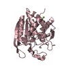
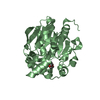
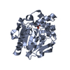
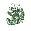
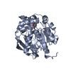
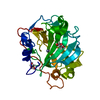
| Entry | Database: PDB / ID: 4g5x | ||||||
|---|---|---|---|---|---|---|---|
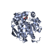
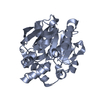
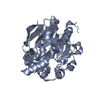
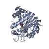
| Title | Crystal structures of N-acyl homoserine lactonase AidH | ||||||
 Components Components | Alpha/beta hydrolase fold protein | ||||||
 Keywords Keywords | HYDROLASE / alpha/beta-hydrolase fold / core domain / eight-stranded sheet / lactonase | ||||||
| Function / homology |  Function and homology information Function and homology information | ||||||
| Biological species |  Ochrobactrum (bacteria) Ochrobactrum (bacteria) | ||||||
| Method |  X-RAY DIFFRACTION / SYNCHROTRON / SAD / Resolution: 1.29 Å X-RAY DIFFRACTION / SYNCHROTRON / SAD / Resolution: 1.29 Å | ||||||
 Authors Authors | Liang, D.C. / Yan, X.X. / Gao, A. | ||||||
 Citation Citation | Journal: Acta Crystallogr.,Sect.D / Year: 2013 Title: High-resolution structures of AidH complexes provide insights into a novel catalytic mechanism for N-acyl homoserine lactonase Authors: Gao, A. / Mei, G.Y. / Liu, S. / Wang, P. / Tang, Q. / Liu, Y.P. / Wen, H. / An, X.M. / Zhang, L.Q. / Yan, X.X. / Liang, D.C. | ||||||
| History |
|
- Structure visualization
| Structure viewer | Molecule: MolmilJmol/JSmol |
|---|
- Downloads & links
-Download
| PDBx/mmCIF format | 4g5x.cif.gz | 266.1 KB | Display | PDBx/mmCIF format |
|---|---|---|---|---|
| PDB format | pdb4g5x.ent.gz | 219.4 KB | Display | PDB format |
| PDBx/mmJSON format | 4g5x.json.gz | Tree view | PDBx/mmJSON format | |
| Others |  Other downloads Other downloads |
-Validation report
| Arichive directory | https://data.pdbj.org/pub/pdb/validation_reports/g5/4g5xftp://data.pdbj.org/pub/pdb/validation_reports/g5/4g5x | HTTPS FTP |
|---|
-Related structure data


-Links
 PDBj
PDBj


- Assembly
| Deposited unit | 
| ||||||||
|---|---|---|---|---|---|---|---|---|---|
| 1 | 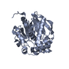
| ||||||||
| 2 | 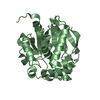
| ||||||||
| Unit cell |
|
-Components
| #1: Protein | Mass: 30653.365 Da / Num. of mol.: 2 Source method: isolated from a genetically manipulated source Source: (gene. exp.) Ochrobactrum (bacteria) / Strain: T63 / Gene: aidH / Production host: #2: Water | ChemComp-HOH / |  Mass: 18.015 Da / Num. of mol.: 903 / Source method: isolated from a natural source / Formula: H2O Mass: 18.015 Da / Num. of mol.: 903 / Source method: isolated from a natural source / Formula: H2O |
|---|
-Experimental details
-Experiment
| Experiment | Method: X-RAY DIFFRACTION / Number of used crystals: 1 |
|---|
- Sample preparation
| Crystal | Density Matthews: 1.87 Å3/Da / Density % sol: 34.24 % |
|---|---|
| Crystal grow | Temperature: 293 K / Method: vapor diffusion, hanging drop / pH: 6.5 Details: pH 6.5, VAPOR DIFFUSION, HANGING DROP, temperature 293K |
-Data collection
| Diffraction | Mean temperature: 180 K |
|---|---|
| Diffraction source | Source: SYNCHROTRON / Site: Photon Factory  / Beamline: BL-17A / Wavelength: 1 Å / Beamline: BL-17A / Wavelength: 1 Å |
| Detector | Type: ADSC QUANTUM 315 / Detector: CCD / Date: Apr 26, 2012 |
| Radiation | Protocol: SINGLE WAVELENGTH / Monochromatic (M) / Laue (L): M / Scattering type: x-ray |
| Radiation wavelength | Wavelength: 1 Å / Relative weight: 1 |
| Reflection | Resolution: 1.29→20 Å / Num. obs: 124835 / % possible obs: 55.3 % / Observed criterion σ(F): 3 / Observed criterion σ(I): 3 / Biso Wilson estimate: 11.42 Å2 |
- Processing
| Software |
| |||||||||||||||||||||||||||||||||||||||||||||||||||||||||||||||||||||||||||||
|---|---|---|---|---|---|---|---|---|---|---|---|---|---|---|---|---|---|---|---|---|---|---|---|---|---|---|---|---|---|---|---|---|---|---|---|---|---|---|---|---|---|---|---|---|---|---|---|---|---|---|---|---|---|---|---|---|---|---|---|---|---|---|---|---|---|---|---|---|---|---|---|---|---|---|---|---|---|---|
| Refinement | Method to determine structure: SAD / Resolution: 1.29→19.87 Å / Occupancy max: 1 / Occupancy min: 0.11 / FOM work R set: 0.9127 / SU ML: 0.15 / σ(F): 0 / Phase error: 15.71 / Stereochemistry target values: ML
| |||||||||||||||||||||||||||||||||||||||||||||||||||||||||||||||||||||||||||||
| Solvent computation | Shrinkage radii: 0.49 Å / VDW probe radii: 0.8 Å / Solvent model: FLAT BULK SOLVENT MODEL / Bsol: 102.138 Å2 / ksol: 0.458 e/Å3 | |||||||||||||||||||||||||||||||||||||||||||||||||||||||||||||||||||||||||||||
| Displacement parameters | Biso max: 94.92 Å2 / Biso mean: 20.9681 Å2 / Biso min: 6.74 Å2
| |||||||||||||||||||||||||||||||||||||||||||||||||||||||||||||||||||||||||||||
| Refinement step | Cycle: LAST / Resolution: 1.29→19.87 Å
| |||||||||||||||||||||||||||||||||||||||||||||||||||||||||||||||||||||||||||||
| Refine LS restraints |
| |||||||||||||||||||||||||||||||||||||||||||||||||||||||||||||||||||||||||||||
| LS refinement shell | Refine-ID: X-RAY DIFFRACTION / Total num. of bins used: 10
|
