 Movie
Movie Controller

[English] 日本語
 Yorodumi
- PDB-4g25: Crystal Structure of proteinaceous RNase P 1 (PRORP1) from A. tha... -
+ Open data

- Basic information
| Entry | Database: PDB / ID: 4g25 | ||||||
|---|---|---|---|---|---|---|---|
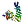
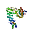
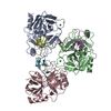
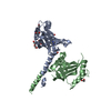
| Title | Crystal Structure of proteinaceous RNase P 1 (PRORP1) from A. thaliana, SeMet substituted form with Sr | ||||||
 Components Components | Pentatricopeptide repeat-containing protein At2g32230, mitochondrial | ||||||
 Keywords Keywords | RNA BINDING PROTEIN / Metallonuclease / PRORP / Ribonuclease / PIN / tRNA processing / Rnase P / NYN domain / PPR domain / Chloroplasts | ||||||
| Function / homology |  Function and homology information Function and homology informationribonuclease P / ribonuclease P activity / tRNA 5'-leader removal / tRNA processing / chloroplast / mitochondrion / metal ion binding Similarity search - Function | ||||||
| Biological species |  | ||||||
| Method |  X-RAY DIFFRACTION / SYNCHROTRON / MOLECULAR REPLACEMENT / Resolution: 2 Å X-RAY DIFFRACTION / SYNCHROTRON / MOLECULAR REPLACEMENT / Resolution: 2 Å | ||||||
 Authors Authors | Koutmos, M. / Howard, M.J. / Fierke, C.A. | ||||||
 Citation Citation | Journal: Proc.Natl.Acad.Sci.USA / Year: 2012 Title: Mitochondrial ribonuclease P structure provides insight into the evolution of catalytic strategies for precursor-tRNA 5' processing. Authors: Howard, M.J. / Lim, W.H. / Fierke, C.A. / Koutmos, M. | ||||||
| History |
|
- Structure visualization
| Structure viewer | Molecule: MolmilJmol/JSmol |
|---|
- Downloads & links
-Download
| PDBx/mmCIF format | 4g25.cif.gz | 117.3 KB | Display | PDBx/mmCIF format |
|---|---|---|---|---|
| PDB format | pdb4g25.ent.gz | 88.8 KB | Display | PDB format |
| PDBx/mmJSON format | 4g25.json.gz | Tree view | PDBx/mmJSON format | |
| Others |  Other downloads Other downloads |
-Validation report
| Summary document | 4g25_validation.pdf.gz | 428.6 KB | Display | wwPDB validaton report |
|---|---|---|---|---|
| Full document | 4g25_full_validation.pdf.gz | 432.7 KB | Display | |
| Data in XML | 4g25_validation.xml.gz | 20.9 KB | Display | |
| Data in CIF | 4g25_validation.cif.gz | 30.9 KB | Display | |
| Arichive directory | https://data.pdbj.org/pub/pdb/validation_reports/g2/4g25ftp://data.pdbj.org/pub/pdb/validation_reports/g2/4g25 | HTTPS FTP |
-Related structure data
| Related structure data |  4g23SC  4g24C  4g26C S: Starting model for refinement C: citing same article ( |
|---|---|
| Similar structure data |


-Links
 PDBj
PDBj

- Assembly
| Deposited unit | 
| ||||||||
|---|---|---|---|---|---|---|---|---|---|
| 1 |
| ||||||||
| Unit cell |
|
-Components
| #1: Protein | Mass: 57317.258 Da / Num. of mol.: 1 / Fragment: UNP residues 77-572 Source method: isolated from a genetically manipulated source Source: (gene. exp.)  |
|---|---|
| #2: Chemical | ChemComp-ZN /   Mass: 65.409 Da / Num. of mol.: 1 / Source method: obtained synthetically / Formula: Zn Mass: 65.409 Da / Num. of mol.: 1 / Source method: obtained synthetically / Formula: Zn |
| #3: Chemical | ChemComp-SR /   Mass: 87.620 Da / Num. of mol.: 1 / Source method: obtained synthetically / Formula: Sr Mass: 87.620 Da / Num. of mol.: 1 / Source method: obtained synthetically / Formula: Sr |
| #4: Water | ChemComp-HOH /  Mass: 18.015 Da / Num. of mol.: 235 / Source method: isolated from a natural source / Formula: H2O Mass: 18.015 Da / Num. of mol.: 235 / Source method: isolated from a natural source / Formula: H2O |
| Has protein modification | Y |
-Experimental details
-Experiment
| Experiment | Method: X-RAY DIFFRACTION / Number of used crystals: 1 |
|---|
- Sample preparation
| Crystal | Density Matthews: 2.86 Å3/Da / Density % sol: 57.02 % |
|---|---|
| Crystal grow | Temperature: 277 K / Method: vapor diffusion, sitting drop / pH: 5.5 Details: 18% PEG 3,350, 0.1 M sodium citrate pH 5.5, and 0.02 M SrCl2, VAPOR DIFFUSION, SITTING DROP, temperature 277K |
-Data collection
| Diffraction | Mean temperature: 100 K |
|---|---|
| Diffraction source | Source: SYNCHROTRON / Site: APS  / Beamline: 23-ID-D / Wavelength: 0.968 Å / Beamline: 23-ID-D / Wavelength: 0.968 Å |
| Detector | Type: MARMOSAIC 300 mm CCD / Detector: CCD / Date: Mar 23, 2012 Details: K-B pair of biomorph mirrors for vertical and horizontal focusing |
| Radiation | Monochromator: double crystal monochromator / Protocol: SINGLE WAVELENGTH / Monochromatic (M) / Laue (L): M / Scattering type: x-ray |
| Radiation wavelength | Wavelength: 0.968 Å / Relative weight: 1 |
| Reflection | Resolution: 2→50 Å / Num. all: 45501 / Num. obs: 44773 / % possible obs: 98.4 % / Observed criterion σ(F): 0 / Observed criterion σ(I): 0 / Redundancy: 5.4 % / Rsym value: 0.084 / Net I/σ(I): 12.6 |
| Reflection shell | Resolution: 2→2.07 Å / Redundancy: 5.4 % / Mean I/σ(I) obs: 2.6 / Num. unique all: 4456 / Rsym value: 0.63 / % possible all: 97.9 |
- Processing
| Software |
| ||||||||||||||||||||||||||||||||||||||||||||||||||||||||||||
|---|---|---|---|---|---|---|---|---|---|---|---|---|---|---|---|---|---|---|---|---|---|---|---|---|---|---|---|---|---|---|---|---|---|---|---|---|---|---|---|---|---|---|---|---|---|---|---|---|---|---|---|---|---|---|---|---|---|---|---|---|---|
| Refinement | Method to determine structure: MOLECULAR REPLACEMENT Starting model: PDB ENTRY 4G23 Resolution: 2→33.5 Å / Cor.coef. Fo:Fc: 0.956 / Cor.coef. Fo:Fc free: 0.939 / SU B: 3.696 / SU ML: 0.103 / Cross valid method: THROUGHOUT / σ(F): 0 / σ(I): 0 / ESU R: 0.157 / ESU R Free: 0.148 / Stereochemistry target values: MAXIMUM LIKELIHOOD / Details: HYDROGENS HAVE BEEN USED IF PRESENT IN THE INPUT
| ||||||||||||||||||||||||||||||||||||||||||||||||||||||||||||
| Solvent computation | Ion probe radii: 0.8 Å / Shrinkage radii: 0.8 Å / VDW probe radii: 1.2 Å / Solvent model: BABINET MODEL WITH MASK | ||||||||||||||||||||||||||||||||||||||||||||||||||||||||||||
| Displacement parameters | Biso mean: 44.137 Å2
| ||||||||||||||||||||||||||||||||||||||||||||||||||||||||||||
| Refinement step | Cycle: LAST / Resolution: 2→33.5 Å
| ||||||||||||||||||||||||||||||||||||||||||||||||||||||||||||
| Refine LS restraints |
| ||||||||||||||||||||||||||||||||||||||||||||||||||||||||||||
| LS refinement shell | Resolution: 2→2.052 Å / Total num. of bins used: 20
|
