 Movie
Movie Controller

[English] 日本語
 Yorodumi
- PDB-6bv9: Structure of proteinaceous RNase P 1 (PRORP1) from A. thaliana af... -
+ Open data

- Basic information
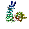
| Entry | Database: PDB / ID: 6bv9 | ||||||
|---|---|---|---|---|---|---|---|
| Title | Structure of proteinaceous RNase P 1 (PRORP1) from A. thaliana after overnight soak with juglone | ||||||
 Components Components | Proteinaceous RNase P 1, chloroplastic/mitochondrial | ||||||
 Keywords Keywords | HYDROLASE/HYDROLASE INHIBITOR / metallonuclease / PRORP / ribonuclease / PIN / tRNA processing / RNAse P / NYN domain / PPR domain / chloroplasts / mitochondria / HYDROLASE-HYDROLASE INHIBITOR complex | ||||||
| Function / homology |  Function and homology information Function and homology informationribonuclease P / ribonuclease P activity / tRNA 5'-leader removal / tRNA processing / chloroplast / mitochondrion / metal ion binding Similarity search - Function | ||||||
| Biological species |  | ||||||
| Method |  X-RAY DIFFRACTION / SYNCHROTRON / MOLECULAR REPLACEMENT / Resolution: 2.1 Å X-RAY DIFFRACTION / SYNCHROTRON / MOLECULAR REPLACEMENT / Resolution: 2.1 Å | ||||||
 Authors Authors | Karasik, A. / Wu, N. / Fierke, C.A. / Koutmos, M. | ||||||
| Funding support |  United States, 1items United States, 1items
| ||||||
 Citation Citation | Journal: To Be Published Title: Inhibition of protein-only RNase P with Gambogic acid and Juglone Authors: Wu, N. / Karasik, A. / Muehlbauer, L. / Koutmos, M. / Fierke, C.A. | ||||||
| History |
|
- Structure visualization
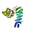
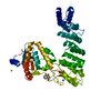
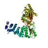
| Structure viewer | Molecule: MolmilJmol/JSmol |
|---|
- Downloads & links
-Download
| PDBx/mmCIF format | 6bv9.cif.gz | 190.8 KB | Display | PDBx/mmCIF format |
|---|---|---|---|---|
| PDB format | pdb6bv9.ent.gz | 153 KB | Display | PDB format |
| PDBx/mmJSON format | 6bv9.json.gz | Tree view | PDBx/mmJSON format | |
| Others |  Other downloads Other downloads |
-Validation report
| Arichive directory | https://data.pdbj.org/pub/pdb/validation_reports/bv/6bv9ftp://data.pdbj.org/pub/pdb/validation_reports/bv/6bv9 | HTTPS FTP |
|---|
-Related structure data
| Related structure data |  6bv5C 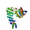 6bv6C 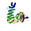 6bv8C 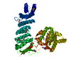 4g23S C: citing same article ( S: Starting model for refinement |
|---|---|
| Similar structure data |


-Links
 PDBj
PDBj
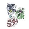
- Assembly
| Deposited unit | 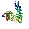
| ||||||||
|---|---|---|---|---|---|---|---|---|---|
| 1 |
| ||||||||
| Unit cell |
|
-Components
| #1: Protein | Mass: 56660.734 Da / Num. of mol.: 1 / Fragment: UNP residues 77-572 Source method: isolated from a genetically manipulated source Source: (gene. exp.)  | ||||||
|---|---|---|---|---|---|---|---|
| #2: Chemical | 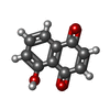  Mass: 174.153 Da / Num. of mol.: 3 / Source method: obtained synthetically / Formula: C10H6O3 Mass: 174.153 Da / Num. of mol.: 3 / Source method: obtained synthetically / Formula: C10H6O3#3: Chemical | ChemComp-ZN / |   Mass: 65.409 Da / Num. of mol.: 1 / Source method: obtained synthetically / Formula: Zn Mass: 65.409 Da / Num. of mol.: 1 / Source method: obtained synthetically / Formula: Zn#4: Chemical | ChemComp-CL / |   Mass: 35.453 Da / Num. of mol.: 1 / Source method: obtained synthetically / Formula: Cl Mass: 35.453 Da / Num. of mol.: 1 / Source method: obtained synthetically / Formula: Cl#5: Water | ChemComp-HOH / |  Mass: 18.015 Da / Num. of mol.: 194 / Source method: isolated from a natural source / Formula: H2O Mass: 18.015 Da / Num. of mol.: 194 / Source method: isolated from a natural source / Formula: H2O |
-Experimental details
-Experiment
| Experiment | Method: X-RAY DIFFRACTION / Number of used crystals: 1 |
|---|
- Sample preparation
| Crystal | Density Matthews: 2.89 Å3/Da / Density % sol: 57.45 % |
|---|---|
| Crystal grow | Temperature: 277 K / Method: vapor diffusion, sitting drop / pH: 5.5 / Details: 18% PEG3350, 0.1 M sodium citrate tribasic, pH 5.5 |
-Data collection
| Diffraction | Mean temperature: 100 K |
|---|---|
| Diffraction source | Source: SYNCHROTRON / Site: APS / Beamline: 23-ID-B / Wavelength: 1.0332 Å |
| Detector | Type: DECTRIS EIGER X 16M / Detector: PIXEL / Date: Feb 8, 2017 Details: K-B pair of biomorph mirrors for vertical and horizontal focusing |
| Radiation | Monochromator: double crystal Si(111) / Protocol: SINGLE WAVELENGTH / Monochromatic (M) / Laue (L): M / Scattering type: x-ray |
| Radiation wavelength | Wavelength: 1.0332 Å / Relative weight: 1 |
| Reflection | Resolution: 2.1→87.21 Å / Num. obs: 39128 / % possible obs: 100 % / Redundancy: 6.7 % / Biso Wilson estimate: 37 Å2 / Rmerge(I) obs: 0.055 / Net I/σ(I): 17.5 |
| Reflection shell | Resolution: 2.1→2.16 Å / Redundancy: 6.5 % / Rmerge(I) obs: 0.717 / Mean I/σ(I) obs: 2.2 / % possible all: 99.7 |
- Processing
| Software |
| ||||||||||||||||||||||||||||||||||||||||||||||||||||||||||||||||||||||||||||||||||||||||||||||||||||||||||||||||||||||||||||||||||||||||||||||||||||||||||||||||||||||||||||||||||||||
|---|---|---|---|---|---|---|---|---|---|---|---|---|---|---|---|---|---|---|---|---|---|---|---|---|---|---|---|---|---|---|---|---|---|---|---|---|---|---|---|---|---|---|---|---|---|---|---|---|---|---|---|---|---|---|---|---|---|---|---|---|---|---|---|---|---|---|---|---|---|---|---|---|---|---|---|---|---|---|---|---|---|---|---|---|---|---|---|---|---|---|---|---|---|---|---|---|---|---|---|---|---|---|---|---|---|---|---|---|---|---|---|---|---|---|---|---|---|---|---|---|---|---|---|---|---|---|---|---|---|---|---|---|---|---|---|---|---|---|---|---|---|---|---|---|---|---|---|---|---|---|---|---|---|---|---|---|---|---|---|---|---|---|---|---|---|---|---|---|---|---|---|---|---|---|---|---|---|---|---|---|---|---|---|
| Refinement | Method to determine structure: MOLECULAR REPLACEMENT Starting model: PDB ENTRY 4G23 Resolution: 2.1→87.21 Å / Cor.coef. Fo:Fc: 0.961 / Cor.coef. Fo:Fc free: 0.937 / SU B: 4.85 / SU ML: 0.127 / Cross valid method: THROUGHOUT / ESU R: 0.183 / ESU R Free: 0.17 / Stereochemistry target values: MAXIMUM LIKELIHOOD / Details: HYDROGENS HAVE BEEN USED IF PRESENT IN THE INPUT
| ||||||||||||||||||||||||||||||||||||||||||||||||||||||||||||||||||||||||||||||||||||||||||||||||||||||||||||||||||||||||||||||||||||||||||||||||||||||||||||||||||||||||||||||||||||||
| Solvent computation | Ion probe radii: 0.8 Å / Shrinkage radii: 0.8 Å / VDW probe radii: 1.2 Å / Solvent model: BABINET MODEL WITH MASK | ||||||||||||||||||||||||||||||||||||||||||||||||||||||||||||||||||||||||||||||||||||||||||||||||||||||||||||||||||||||||||||||||||||||||||||||||||||||||||||||||||||||||||||||||||||||
| Displacement parameters | Biso mean: 63.12 Å2
| ||||||||||||||||||||||||||||||||||||||||||||||||||||||||||||||||||||||||||||||||||||||||||||||||||||||||||||||||||||||||||||||||||||||||||||||||||||||||||||||||||||||||||||||||||||||
| Refinement step | Cycle: LAST / Resolution: 2.1→87.21 Å
| ||||||||||||||||||||||||||||||||||||||||||||||||||||||||||||||||||||||||||||||||||||||||||||||||||||||||||||||||||||||||||||||||||||||||||||||||||||||||||||||||||||||||||||||||||||||
| Refine LS restraints |
|