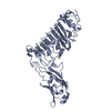
- PDB-4fvf: SPFH domain of mouse stomatin (Crystal form 1) -
+
Open data
ID or keywords:
Loading...
-
Basic information
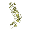
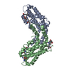
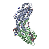
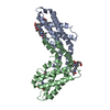
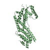
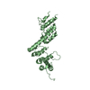
Entry
Database: PDB / ID: 4fvf
Title
SPFH domain of mouse stomatin (Crystal form 1)
Components
Stomatin
Keywords
MEMBRANE PROTEIN / mixed alpha-beta / scaffolding protein
Function / homology
Function and homology information
RHOH GTPase cycle / RHOQ GTPase cycle / positive regulation of viral process / RHOC GTPase cycle / RHOB GTPase cycle / RHOA GTPase cycle / host-mediated activation of viral genome replication / Stimuli-sensing channels / regulation of monoatomic ion transmembrane transport / RNA polymerase binding ...RHOH GTPase cycle / RHOQ GTPase cycle / positive regulation of viral process / RHOC GTPase cycle / RHOB GTPase cycle / RHOA GTPase cycle / host-mediated activation of viral genome replication / Stimuli-sensing channels / regulation of monoatomic ion transmembrane transport / RNA polymerase binding / ion channel inhibitor activity / positive regulation of protein targeting to membrane / Neutrophil degranulation / melanosome / cytoskeleton / membrane raft / perinuclear region of cytoplasm / endoplasmic reticulum / protein homodimerization activity / mitochondrion / identical protein binding / plasma membrane / cytosol / cytoplasm Similarity search - Function
Band 7 domain / Band-7 stomatin-like / Band 7/stomatin-like, conserved site / Band 7 protein family signature. / Stomatin/HflK/HflC family / Tetrahydropterin Synthase; Chain A / Band 7 domain / SPFH domain / Band 7 family / prohibitin homologues / Band 7/SPFH domain superfamily ...Band 7 domain / Band-7 stomatin-like / Band 7/stomatin-like, conserved site / Band 7 protein family signature. / Stomatin/HflK/HflC family / Tetrahydropterin Synthase; Chain A / Band 7 domain / SPFH domain / Band 7 family / prohibitin homologues / Band 7/SPFH domain superfamily / 2-Layer Sandwich / Alpha Beta Similarity search - Domain/homology
In the structure databanks used in Yorodumi, some data are registered as the other names, "COVID-19 virus" and "2019-nCoV". Here are the details of the virus and the list of structure data.
Jan 31, 2019. EMDB accession codes are about to change! (news from PDBe EMDB page)
EMDB accession codes are about to change! (news from PDBe EMDB page)
The allocation of 4 digits for EMDB accession codes will soon come to an end. Whilst these codes will remain in use, new EMDB accession codes will include an additional digit and will expand incrementally as the available range of codes is exhausted. The current 4-digit format prefixed with “EMD-” (i.e. EMD-XXXX) will advance to a 5-digit format (i.e. EMD-XXXXX), and so on. It is currently estimated that the 4-digit codes will be depleted around Spring 2019, at which point the 5-digit format will come into force.
The EM Navigator/Yorodumi systems omit the EMD- prefix.
Related info.:Q: What is EMD? / ID/Accession-code notation in Yorodumi/EM Navigator
Yorodumi is a browser for structure data from EMDB, PDB, SASBDB, etc.
This page is also the successor to EM Navigator detail page, and also detail information page/front-end page for Omokage search.
The word "yorodu" (or yorozu) is an old Japanese word meaning "ten thousand". "mi" (miru) is to see.
Related info.:EMDB / PDB / SASBDB / Comparison of 3 databanks / Yorodumi Search / Aug 31, 2016. New EM Navigator & Yorodumi / Yorodumi Papers / Jmol/JSmol / Function and homology information / Changes in new EM Navigator and Yorodumi
 Movie
Movie Controller


 Open data
 Basic information
 Components
 Keywords
 Function and homology information
 X-RAY DIFFRACTION /
  Authors
 Citation
 Structure visualization
 Downloads & links
 Other downloads
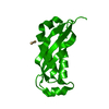


 PDBj
PDBj


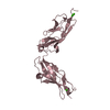
 Assembly
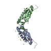


 Mass: 112.411 Da / Num. of mol.: 13 / Source method: obtained synthetically / Formula: Cd
 Mass: 59.044 Da / Num. of mol.: 1 / Source method: obtained synthetically / Formula: C2H3O2
 Mass: 18.015 Da / Num. of mol.: 77 / Source method: isolated from a natural source / Formula: H2O
 Sample preparation
 / Beamline: X06SA / Wavelength: 0.91841 Å
 Processing