 Movie
Movie Controller

[English] 日本語
 Yorodumi
- PDB-4fvd: Crystal structure of EV71 2A proteinase C110A mutant in complex w... -
+ Open data

- Basic information
| Entry | Database: PDB / ID: 4fvd | ||||||
|---|---|---|---|---|---|---|---|
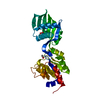
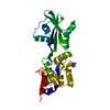
| Title | Crystal structure of EV71 2A proteinase C110A mutant in complex with substrate | ||||||
 Components Components |
| ||||||
 Keywords Keywords | HYDROLASE/HYDROLASE SUBSTRATE / HYDROLASE / CYSTEINE PROTEINASE / HYDROLASE-HYDROLASE SUBSTRATE complex | ||||||
| Function / homology |  Function and homology information Function and homology informationsymbiont-mediated suppression of host cytoplasmic pattern recognition receptor signaling pathway via inhibition of MDA-5 activity / picornain 2A / symbiont-mediated suppression of host mRNA export from nucleus / symbiont genome entry into host cell via pore formation in plasma membrane / picornain 3C / T=pseudo3 icosahedral viral capsid / ribonucleoside triphosphate phosphatase activity / host cell cytoplasmic vesicle membrane / nucleoside-triphosphate phosphatase / channel activity ...symbiont-mediated suppression of host cytoplasmic pattern recognition receptor signaling pathway via inhibition of MDA-5 activity / picornain 2A / symbiont-mediated suppression of host mRNA export from nucleus / symbiont genome entry into host cell via pore formation in plasma membrane / picornain 3C / T=pseudo3 icosahedral viral capsid / ribonucleoside triphosphate phosphatase activity / host cell cytoplasmic vesicle membrane / nucleoside-triphosphate phosphatase / channel activity / monoatomic ion transmembrane transport / DNA replication / RNA helicase activity / endocytosis involved in viral entry into host cell / symbiont-mediated activation of host autophagy / RNA-directed RNA polymerase / cysteine-type endopeptidase activity / viral RNA genome replication / RNA-directed RNA polymerase activity / DNA-templated transcription / virion attachment to host cell / host cell nucleus / structural molecule activity / proteolysis / RNA binding / zinc ion binding / ATP binding / membrane Similarity search - Function | ||||||
| Biological species |   Human enterovirus 71 Human enterovirus 71 | ||||||
| Method |  X-RAY DIFFRACTION / MOLECULAR REPLACEMENT / molecular replacement / Resolution: 1.66 Å X-RAY DIFFRACTION / MOLECULAR REPLACEMENT / molecular replacement / Resolution: 1.66 Å | ||||||
 Authors Authors | Cai, Q. / Muhammad, Y. / Liu, W. / Gao, Z. / Peng, X. / Cai, Y. / Wu, C. / Zheng, Q. / Li, J. / Lin, T. | ||||||
 Citation Citation | Journal: J.Virol. / Year: 2013 Title: Conformational Plasticity of 2A Proteinase from Enterovirus 71 Authors: Cai, Q. / Yameen, M. / Liu, W. / Gao, Z. / Li, Y. / Peng, X. / Cai, Y. / Wu, C. / Zheng, Q. / Li, J. / Lin, T. | ||||||
| History |
|
- Structure visualization
| Structure viewer | Molecule: MolmilJmol/JSmol |
|---|
- Downloads & links
-Download
| PDBx/mmCIF format | 4fvd.cif.gz | 45.1 KB | Display | PDBx/mmCIF format |
|---|---|---|---|---|
| PDB format | pdb4fvd.ent.gz | 30.5 KB | Display | PDB format |
| PDBx/mmJSON format | 4fvd.json.gz | Tree view | PDBx/mmJSON format | |
| Others |  Other downloads Other downloads |
-Validation report
| Arichive directory | https://data.pdbj.org/pub/pdb/validation_reports/fv/4fvdftp://data.pdbj.org/pub/pdb/validation_reports/fv/4fvd | HTTPS FTP |
|---|
-Related structure data
| Related structure data |  4fvbSC  4fve 4fvh 4fvi S: Starting model for refinement C: citing same article ( |
|---|---|
| Similar structure data |


-Links
 PDBj
PDBj

- Assembly
| Deposited unit | 
| ||||||||
|---|---|---|---|---|---|---|---|---|---|
| 1 | 
| ||||||||
| Unit cell |
| ||||||||
| Components on special symmetry positions |
|
-Components
| #1: Protein | Mass: 16706.549 Da / Num. of mol.: 1 / Mutation: C110A Source method: isolated from a genetically manipulated source Source: (gene. exp.) Human enterovirus 71 / Strain: E2004104-TW-CDC / Gene: polyprotein / Plasmid: pGEX-4T-1 / Production host:  |
|---|---|
| #2: Protein/peptide | Mass: 981.125 Da / Num. of mol.: 1 Source method: isolated from a genetically manipulated source Source: (gene. exp.) Human enterovirus 71 / Strain: E2004104-TW-CDC / Gene: polyprotein / Plasmid: pGEX-4T-1 / Production host: |
| #3: Chemical | ChemComp-ZN /   Mass: 65.409 Da / Num. of mol.: 1 / Source method: obtained synthetically / Formula: Zn Mass: 65.409 Da / Num. of mol.: 1 / Source method: obtained synthetically / Formula: Zn |
| #4: Water | ChemComp-HOH /  Mass: 18.015 Da / Num. of mol.: 133 / Source method: isolated from a natural source / Formula: H2O Mass: 18.015 Da / Num. of mol.: 133 / Source method: isolated from a natural source / Formula: H2O |
| Has protein modification | Y |
-Experimental details
-Experiment
| Experiment | Method: X-RAY DIFFRACTION / Number of used crystals: 1 |
|---|
- Sample preparation
| Crystal | Density Matthews: 2.58 Å3/Da / Density % sol: 52.32 % |
|---|---|
| Crystal grow | Temperature: 289 K / Method: vapor diffusion, hanging drop / pH: 7.5 Details: 100mM HEPES, 20% 2-propanol, 10% PEG 4000, pH 7.5, vapor diffusion, hanging drop, temperature 289K |
-Data collection
| Diffraction | Mean temperature: 100 K |
|---|---|
| Diffraction source | Source: ROTATING ANODE / Type: RIGAKU MICROMAX-007 HF / Wavelength: 1.5418 Å |
| Detector | Type: MAR scanner 345 mm plate / Detector: IMAGE PLATE / Date: May 27, 2010 |
| Radiation | Protocol: SINGLE WAVELENGTH / Monochromatic (M) / Laue (L): M / Scattering type: x-ray |
| Radiation wavelength | Wavelength: 1.5418 Å / Relative weight: 1 |
| Reflection | Resolution: 1.66→50 Å / Num. all: 21456 / Num. obs: 20028 / % possible obs: 93.3 % / Observed criterion σ(F): 0 / Observed criterion σ(I): 0 / Redundancy: 3.66 % / Rmerge(I) obs: 0.0386 / Net I/σ(I): 11.5 |
| Reflection shell | Resolution: 1.66→1.72 Å / Redundancy: 3.29 % / Rmerge(I) obs: 0.4443 / Mean I/σ(I) obs: 1.2 / % possible all: 78.3 |
-Phasing
| Phasing | Method: molecular replacement |
|---|
- Processing
| Software |
| |||||||||||||||||||||||||||||||||||||||||||||||||||||||||||||||||
|---|---|---|---|---|---|---|---|---|---|---|---|---|---|---|---|---|---|---|---|---|---|---|---|---|---|---|---|---|---|---|---|---|---|---|---|---|---|---|---|---|---|---|---|---|---|---|---|---|---|---|---|---|---|---|---|---|---|---|---|---|---|---|---|---|---|---|
| Refinement | Method to determine structure: MOLECULAR REPLACEMENT Starting model: 4FVB Resolution: 1.66→40.07 Å / Cor.coef. Fo:Fc: 0.964 / Cor.coef. Fo:Fc free: 0.948 / Occupancy max: 1 / Occupancy min: 0.5 / SU B: 1.993 / SU ML: 0.067 / Cross valid method: THROUGHOUT / σ(F): 0 / ESU R: 0.097 / ESU R Free: 0.1 / Stereochemistry target values: MAXIMUM LIKELIHOOD Details: HYDROGENS HAVE BEEN ADDED IN THE RIDING POSITIONS U VALUES: REFINED INDIVIDUALLY
| |||||||||||||||||||||||||||||||||||||||||||||||||||||||||||||||||
| Solvent computation | Ion probe radii: 0.8 Å / Shrinkage radii: 0.8 Å / VDW probe radii: 1.4 Å / Solvent model: MASK | |||||||||||||||||||||||||||||||||||||||||||||||||||||||||||||||||
| Displacement parameters | Biso max: 67.21 Å2 / Biso mean: 27.4618 Å2 / Biso min: 11.72 Å2
| |||||||||||||||||||||||||||||||||||||||||||||||||||||||||||||||||
| Refinement step | Cycle: LAST / Resolution: 1.66→40.07 Å
| |||||||||||||||||||||||||||||||||||||||||||||||||||||||||||||||||
| Refine LS restraints |
| |||||||||||||||||||||||||||||||||||||||||||||||||||||||||||||||||
| LS refinement shell | Resolution: 1.661→1.704 Å / Total num. of bins used: 20
|
