 Movie
Movie Controller

[English] 日本語
 Yorodumi
- PDB-4ftw: Crystal structure of a carboxyl esterase N110C/L145H at 2.3 angst... -
+ Open data

- Basic information
| Entry | Database: PDB / ID: 4ftw | ||||||
|---|---|---|---|---|---|---|---|
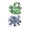
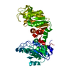
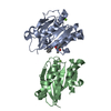
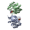
| Title | Crystal structure of a carboxyl esterase N110C/L145H at 2.3 angstrom resolution | ||||||
 Components Components | Phospholipase/Carboxylesterase | ||||||
 Keywords Keywords | HYDROLASE / alpha/beta hydrolase superfamily / esterase | ||||||
| Function / homology |  Function and homology information Function and homology information | ||||||
| Biological species |  Rhodobacter sphaeroides (bacteria) Rhodobacter sphaeroides (bacteria) | ||||||
| Method |  X-RAY DIFFRACTION / SYNCHROTRON / MOLECULAR REPLACEMENT / Resolution: 2.3 Å X-RAY DIFFRACTION / SYNCHROTRON / MOLECULAR REPLACEMENT / Resolution: 2.3 Å | ||||||
 Authors Authors | Wu, L. / Ma, J. / Zhou, J. / Yu, H. | ||||||
 Citation Citation | Journal: Appl.Microbiol.Biotechnol. / Year: 2013 Title: Enhanced enantioselectivity of a carboxyl esterase from Rhodobacter sphaeroides by directed evolution. Authors: Ma, J. / Wu, L. / Guo, F. / Gu, J. / Tang, X. / Jiang, L. / Liu, J. / Zhou, J. / Yu, H. | ||||||
| History |
|
- Structure visualization
| Structure viewer | Molecule: MolmilJmol/JSmol |
|---|
- Downloads & links
-Download
| PDBx/mmCIF format | 4ftw.cif.gz | 58.3 KB | Display | PDBx/mmCIF format |
|---|---|---|---|---|
| PDB format | pdb4ftw.ent.gz | 40.4 KB | Display | PDB format |
| PDBx/mmJSON format | 4ftw.json.gz | Tree view | PDBx/mmJSON format | |
| Others |  Other downloads Other downloads |
-Validation report
| Summary document | 4ftw_validation.pdf.gz | 1007.2 KB | Display | wwPDB validaton report |
|---|---|---|---|---|
| Full document | 4ftw_full_validation.pdf.gz | 1015.1 KB | Display | |
| Data in XML | 4ftw_validation.xml.gz | 12.5 KB | Display | |
| Data in CIF | 4ftw_validation.cif.gz | 15.7 KB | Display | |
| Arichive directory | https://data.pdbj.org/pub/pdb/validation_reports/ft/4ftwftp://data.pdbj.org/pub/pdb/validation_reports/ft/4ftw | HTTPS FTP |
-Related structure data
| Related structure data |  4fhzSC S: Starting model for refinement C: citing same article ( |
|---|---|
| Similar structure data |

-Links
 PDBj
PDBj- Assembly
| Deposited unit | 
| ||||||||
|---|---|---|---|---|---|---|---|---|---|
| 1 | 
| ||||||||
| Unit cell |
|
-Components
-Protein / Sugars , 2 types, 2 molecules A
| #1: Protein | Mass: 29919.902 Da / Num. of mol.: 1 / Mutation: N110C/L145H Source method: isolated from a genetically manipulated source Source: (gene. exp.) Rhodobacter sphaeroides (bacteria) / Strain: ATCC 17023 / 2.4.1 / NCIB 8253 / DSM 158 / Gene: CGMCC1.1737, RHOS4_13150, RSP_2728 / Plasmid: pET-30a / Production host: |
|---|---|
| #3: Sugar | ChemComp-3CM /  Type: D-saccharide / Mass: 466.520 Da / Num. of mol.: 1 Type: D-saccharide / Mass: 466.520 Da / Num. of mol.: 1Source method: isolated from a genetically manipulated source Formula: C21H38O11 |
-Non-polymers , 4 types, 25 molecules 


| #2: Chemical | ChemComp-PIN /  Mass: 302.368 Da / Num. of mol.: 1 / Source method: obtained synthetically / Formula: C8H18N2O6S2 / Comment: pH buffer*YM Mass: 302.368 Da / Num. of mol.: 1 / Source method: obtained synthetically / Formula: C8H18N2O6S2 / Comment: pH buffer*YM |
|---|---|
| #4: Chemical | ChemComp-NA /  Mass: 22.990 Da / Num. of mol.: 1 / Source method: obtained synthetically / Formula: Na Mass: 22.990 Da / Num. of mol.: 1 / Source method: obtained synthetically / Formula: Na |
| #5: Chemical | ChemComp-CL /  Mass: 35.453 Da / Num. of mol.: 1 / Source method: obtained synthetically / Formula: Cl Mass: 35.453 Da / Num. of mol.: 1 / Source method: obtained synthetically / Formula: Cl |
| #6: Water | ChemComp-HOH / Mass: 18.015 Da / Num. of mol.: 22 / Source method: isolated from a natural source / Formula: H2O |
-Details
| Has protein modification | Y |
|---|
-Experimental details
-Experiment
| Experiment | Method: X-RAY DIFFRACTION / Number of used crystals: 1 |
|---|
- Sample preparation
| Crystal | Density Matthews: 3.49 Å3/Da / Density % sol: 64.78 % |
|---|---|
| Crystal grow | Temperature: 293 K / Method: vapor diffusion, sitting drop / pH: 5.5 Details: 1.3M Na-tartrate, 34.5mM CYMAL-3, 0.1M pipes , pH 5.5, VAPOR DIFFUSION, SITTING DROP, temperature 293K |
-Data collection
| Diffraction | Mean temperature: 100 K | ||||||||||||||||||||||||||||
|---|---|---|---|---|---|---|---|---|---|---|---|---|---|---|---|---|---|---|---|---|---|---|---|---|---|---|---|---|---|
| Diffraction source | Source: SYNCHROTRON / Site: SSRF  / Beamline: BL17U / Wavelength: 0.979 Å / Beamline: BL17U / Wavelength: 0.979 Å | ||||||||||||||||||||||||||||
| Detector | Type: ADSC QUANTUM 315 / Detector: CCD / Date: 2012 | ||||||||||||||||||||||||||||
| Radiation | Monochromator: Si 111 CHANNEL / Protocol: SINGLE WAVELENGTH / Monochromatic (M) / Laue (L): M / Scattering type: x-ray | ||||||||||||||||||||||||||||
| Radiation wavelength | Wavelength: 0.979 Å / Relative weight: 1 | ||||||||||||||||||||||||||||
| Reflection | Resolution: 2.3→50 Å / Num. obs: 15208 / % possible obs: 97.4 % / Observed criterion σ(F): 0 / Observed criterion σ(I): -3 / Redundancy: 9.8 % / Rmerge(I) obs: 0.088 / Rsym value: 0.088 / Net I/σ(I): 20.538 | ||||||||||||||||||||||||||||
| Reflection shell |
|
- Processing
| Software |
| ||||||||||||||||||||||||||||||||||||||||||
|---|---|---|---|---|---|---|---|---|---|---|---|---|---|---|---|---|---|---|---|---|---|---|---|---|---|---|---|---|---|---|---|---|---|---|---|---|---|---|---|---|---|---|---|
| Refinement | Method to determine structure: MOLECULAR REPLACEMENT Starting model: PDB ENTRY 4FHZ Resolution: 2.3→48.663 Å / SU ML: 0.35 / Isotropic thermal model: Isotropic / σ(F): 0 / Phase error: 26.99 / Stereochemistry target values: ML
| ||||||||||||||||||||||||||||||||||||||||||
| Solvent computation | Shrinkage radii: 0.49 Å / VDW probe radii: 0.8 Å / Solvent model: FLAT BULK SOLVENT MODEL / Bsol: 72.139 Å2 / ksol: 0.387 e/Å3 | ||||||||||||||||||||||||||||||||||||||||||
| Displacement parameters |
| ||||||||||||||||||||||||||||||||||||||||||
| Refinement step | Cycle: LAST / Resolution: 2.3→48.663 Å
| ||||||||||||||||||||||||||||||||||||||||||
| Refine LS restraints |
| ||||||||||||||||||||||||||||||||||||||||||
| LS refinement shell |
|
