 Movie
Movie Controller

[English] 日本語
 Yorodumi
- PDB-4fpy: Crystal structure of the NanB sialidase from streptococcus pneumo... -
+ Open data

- Basic information
| Entry | Database: PDB / ID: 4fpy | ||||||
|---|---|---|---|---|---|---|---|
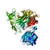
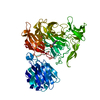
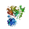
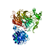
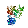
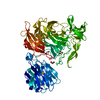
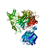
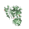
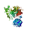
| Title | Crystal structure of the NanB sialidase from streptococcus pneumoniae in complex with 2-[(3-Bromobenzyl)ammonio]ethanesulfonate | ||||||
 Components Components | Sialidase B | ||||||
 Keywords Keywords | HYDROLASE/INHIBITOR / hydrolase / intramolecular trans-sialidase / glycosidase / drug design / neuraminidase / HYDROLASE-INHIBITOR complex | ||||||
| Function / homology |  Function and homology information Function and homology informationganglioside catabolic process / oligosaccharide catabolic process / exo-alpha-sialidase / exo-alpha-sialidase activity / membrane / cytoplasm Similarity search - Function | ||||||
| Biological species |   Streptococcus pneumoniae (bacteria) Streptococcus pneumoniae (bacteria) | ||||||
| Method |  X-RAY DIFFRACTION / MOLECULAR REPLACEMENT / Resolution: 2.18 Å X-RAY DIFFRACTION / MOLECULAR REPLACEMENT / Resolution: 2.18 Å | ||||||
 Authors Authors | Brear, P. | ||||||
 Citation Citation | Journal: Chembiochem / Year: 2012 Title: Synthesis and structural characterisation of selective non-carbohydrate-based inhibitors of bacterial sialidases. Authors: Brear, P. / Telford, J. / Taylor, G.L. / Westwood, N.J. | ||||||
| History |
|
- Structure visualization
| Structure viewer | Molecule: MolmilJmol/JSmol |
|---|
- Downloads & links
-Download
| PDBx/mmCIF format | 4fpy.cif.gz | 153.1 KB | Display | PDBx/mmCIF format |
|---|---|---|---|---|
| PDB format | pdb4fpy.ent.gz | 117.7 KB | Display | PDB format |
| PDBx/mmJSON format | 4fpy.json.gz | Tree view | PDBx/mmJSON format | |
| Others |  Other downloads Other downloads |
-Validation report
| Arichive directory | https://data.pdbj.org/pub/pdb/validation_reports/fp/4fpyftp://data.pdbj.org/pub/pdb/validation_reports/fp/4fpy | HTTPS FTP |
|---|
-Related structure data
| Related structure data |  4foqC  4fovC  4fowC 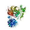 4foyC  4fp2C 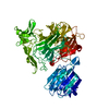 4fp3C 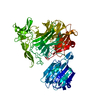 4fpcC 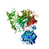 4fpeC 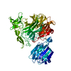 4fpfC 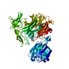 4fpgC 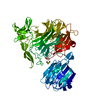 4fphC 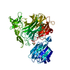 4fpjC  4fpkC  4fplC  4fpoC  4fq4C C: citing same article ( |
|---|---|
| Similar structure data |


-Links
 PDBj
PDBj

- Assembly
| Deposited unit | 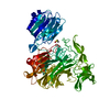
| ||||||||
|---|---|---|---|---|---|---|---|---|---|
| 1 |
| ||||||||
| Unit cell |
|
-Components
| #1: Protein | Mass: 77780.883 Da / Num. of mol.: 1 Source method: isolated from a genetically manipulated source Source: (gene. exp.) Streptococcus pneumoniae (bacteria) / Strain: TIGR4 / Gene: nanB, SP_1687 / Plasmid: pET23b / Production host: | ||
|---|---|---|---|
| #2: Chemical | ChemComp-0V8 /   Mass: 294.165 Da / Num. of mol.: 1 / Source method: obtained synthetically / Formula: C9H12BrNO3S Mass: 294.165 Da / Num. of mol.: 1 / Source method: obtained synthetically / Formula: C9H12BrNO3S | ||
| #3: Chemical |   Mass: 78.133 Da / Num. of mol.: 2 / Source method: obtained synthetically / Formula: C2H6OS / Comment: DMSO, precipitant*YM Mass: 78.133 Da / Num. of mol.: 2 / Source method: obtained synthetically / Formula: C2H6OS / Comment: DMSO, precipitant*YM#4: Water | ChemComp-HOH / |  Mass: 18.015 Da / Num. of mol.: 443 / Source method: isolated from a natural source / Formula: H2O Mass: 18.015 Da / Num. of mol.: 443 / Source method: isolated from a natural source / Formula: H2O |
-Experimental details
-Experiment
| Experiment | Method: X-RAY DIFFRACTION / Number of used crystals: 1 |
|---|
- Sample preparation
| Crystal | Density Matthews: 2.34 Å3/Da / Density % sol: 47.49 % |
|---|---|
| Crystal grow | Temperature: 298 K / Method: vapor diffusion, sitting drop / pH: 8 Details: 7% PEG 8000, 0.1M IMIDAZOLE, pH 8.0, vapor diffusion, sitting drop, temperature 298K |
-Data collection
| Diffraction | Mean temperature: 100 K | |||||||||||||||||||||||||||||||||||||||||||||||||||||||||||||||||||||||||||||||||||||||||||||||||||||||||||||||||||||||||||||||||||||||||||||||||||
|---|---|---|---|---|---|---|---|---|---|---|---|---|---|---|---|---|---|---|---|---|---|---|---|---|---|---|---|---|---|---|---|---|---|---|---|---|---|---|---|---|---|---|---|---|---|---|---|---|---|---|---|---|---|---|---|---|---|---|---|---|---|---|---|---|---|---|---|---|---|---|---|---|---|---|---|---|---|---|---|---|---|---|---|---|---|---|---|---|---|---|---|---|---|---|---|---|---|---|---|---|---|---|---|---|---|---|---|---|---|---|---|---|---|---|---|---|---|---|---|---|---|---|---|---|---|---|---|---|---|---|---|---|---|---|---|---|---|---|---|---|---|---|---|---|---|---|---|---|
| Diffraction source | Source: ROTATING ANODE / Type: RIGAKU MICROMAX-007 HF / Wavelength: 1.5418 Å | |||||||||||||||||||||||||||||||||||||||||||||||||||||||||||||||||||||||||||||||||||||||||||||||||||||||||||||||||||||||||||||||||||||||||||||||||||
| Detector | Type: RIGAKU SATURN 944 / Detector: CCD / Date: Sep 13, 2011 | |||||||||||||||||||||||||||||||||||||||||||||||||||||||||||||||||||||||||||||||||||||||||||||||||||||||||||||||||||||||||||||||||||||||||||||||||||
| Radiation | Protocol: SINGLE WAVELENGTH / Monochromatic (M) / Laue (L): M / Scattering type: x-ray | |||||||||||||||||||||||||||||||||||||||||||||||||||||||||||||||||||||||||||||||||||||||||||||||||||||||||||||||||||||||||||||||||||||||||||||||||||
| Radiation wavelength | Wavelength: 1.5418 Å / Relative weight: 1 | |||||||||||||||||||||||||||||||||||||||||||||||||||||||||||||||||||||||||||||||||||||||||||||||||||||||||||||||||||||||||||||||||||||||||||||||||||
| Reflection | Resolution: 2.18→30 Å / Num. obs: 36283 / % possible obs: 93.3 % / Redundancy: 5.1 % / Rmerge(I) obs: 0.07 / Χ2: 2.244 / Net I/σ(I): 15.7 | |||||||||||||||||||||||||||||||||||||||||||||||||||||||||||||||||||||||||||||||||||||||||||||||||||||||||||||||||||||||||||||||||||||||||||||||||||
| Reflection shell |
|
- Processing
| Software |
| ||||||||||||||||||||||||||||
|---|---|---|---|---|---|---|---|---|---|---|---|---|---|---|---|---|---|---|---|---|---|---|---|---|---|---|---|---|---|
| Refinement | Method to determine structure: MOLECULAR REPLACEMENT / Resolution: 2.18→21.268 Å / Occupancy max: 1 / Occupancy min: 0.42 / SU ML: 0.29 / σ(F): 1.34 / Phase error: 23.53 / Stereochemistry target values: ML
| ||||||||||||||||||||||||||||
| Solvent computation | Shrinkage radii: 0.86 Å / VDW probe radii: 1.1 Å / Solvent model: FLAT BULK SOLVENT MODEL / Bsol: 51.599 Å2 / ksol: 0.358 e/Å3 | ||||||||||||||||||||||||||||
| Displacement parameters | Biso max: 101.12 Å2 / Biso mean: 24.634 Å2 / Biso min: 8.28 Å2
| ||||||||||||||||||||||||||||
| Refinement step | Cycle: LAST / Resolution: 2.18→21.268 Å
|
