[English] 日本語
Yorodumi
- PDB-4flb: CID of human RPRD2 -

+
Open data


ID or keywords:

Loading...

-
Basic information

Entry
Database: PDB / ID: 4flb
TitleCID of human RPRD2
ComponentsRegulation of nuclear pre-mRNA domain-containing protein 2
KeywordsPROTEIN BINDING / structural genomics consortium / sgc
Function / homology
Function and homology information


RNA polymerase II, holoenzyme / snRNA transcription by RNA polymerase II / mRNA 3'-end processing / RNA polymerase II complex binding / RNA polymerase II transcribes snRNA genes / nucleoplasm
Similarity search - Function
Regulation of nuclear pre-mRNA domain-containing protein 1A/B / Cell-cycle alteration and expression-elevated protein in tumour / CID domain / RPR / CID domain / CID domain profile. / Serine Threonine Protein Phosphatase 5, Tetratricopeptide repeat - #90 / ENTH/VHS / Serine Threonine Protein Phosphatase 5, Tetratricopeptide repeat / Alpha Horseshoe / Mainly Alpha
Similarity search - Domain/homology
PRASEODYMIUM ION / Regulation of nuclear pre-mRNA domain-containing protein 2
Similarity search - Component
Biological speciesHomo sapiens (human)
MethodX-RAY DIFFRACTION / SAD / Resolution: 1.802 Å
AuthorsNi, Z. / Xu, C. / Tempel, W. / El Bakkouri, M. / Loppnau, P. / Guo, X. / Bountra, C. / Arrowsmith, C.H. / Edwards, A.M. / Min, J. ...Ni, Z. / Xu, C. / Tempel, W. / El Bakkouri, M. / Loppnau, P. / Guo, X. / Bountra, C. / Arrowsmith, C.H. / Edwards, A.M. / Min, J. / Greenblatt, J.F. / Structural Genomics Consortium (SGC)
CitationJournal: Nat.Struct.Mol.Biol. / Year: 2014
Title: RPRD1A and RPRD1B are human RNA polymerase II C-terminal domain scaffolds for Ser5 dephosphorylation.
Authors: Ni, Z. / Xu, C. / Guo, X. / Hunter, G.O. / Kuznetsova, O.V. / Tempel, W. / Marcon, E. / Zhong, G. / Guo, H. / Kuo, W.H. / Li, J. / Young, P. / Olsen, J.B. / Wan, C. / Loppnau, P. / El ...Authors: Ni, Z. / Xu, C. / Guo, X. / Hunter, G.O. / Kuznetsova, O.V. / Tempel, W. / Marcon, E. / Zhong, G. / Guo, H. / Kuo, W.H. / Li, J. / Young, P. / Olsen, J.B. / Wan, C. / Loppnau, P. / El Bakkouri, M. / Senisterra, G.A. / He, H. / Huang, H. / Sidhu, S.S. / Emili, A. / Murphy, S. / Mosley, A.L. / Arrowsmith, C.H. / Min, J. / Greenblatt, J.F.
History
DepositionJun 14, 2012Deposition site: RCSB / Processing site: RCSB
Revision 1.0Aug 15, 2012Provider: repository / Type: Initial release
Revision 1.1Jun 11, 2014Group: Structure summary
Revision 1.2Jul 30, 2014Group: Database references
Revision 1.3Aug 20, 2014Group: Database references
Revision 1.4Feb 28, 2024Group: Data collection / Database references / Derived calculations
Category: chem_comp_atom / chem_comp_bond ...chem_comp_atom / chem_comp_bond / database_2 / diffrn_source / pdbx_struct_conn_angle / struct_conn / struct_ref_seq_dif / struct_site
Item: _database_2.pdbx_DOI / _database_2.pdbx_database_accession ..._database_2.pdbx_DOI / _database_2.pdbx_database_accession / _diffrn_source.type / _pdbx_struct_conn_angle.ptnr1_auth_comp_id / _pdbx_struct_conn_angle.ptnr1_auth_seq_id / _pdbx_struct_conn_angle.ptnr1_label_atom_id / _pdbx_struct_conn_angle.ptnr1_label_comp_id / _pdbx_struct_conn_angle.ptnr1_label_seq_id / _pdbx_struct_conn_angle.ptnr3_auth_comp_id / _pdbx_struct_conn_angle.ptnr3_auth_seq_id / _pdbx_struct_conn_angle.ptnr3_label_atom_id / _pdbx_struct_conn_angle.ptnr3_label_comp_id / _pdbx_struct_conn_angle.ptnr3_label_seq_id / _pdbx_struct_conn_angle.value / _struct_conn.pdbx_dist_value / _struct_conn.ptnr1_auth_comp_id / _struct_conn.ptnr1_auth_seq_id / _struct_conn.ptnr1_label_atom_id / _struct_conn.ptnr1_label_comp_id / _struct_conn.ptnr1_label_seq_id / _struct_conn.ptnr2_auth_seq_id / _struct_conn.ptnr2_label_asym_id / _struct_ref_seq_dif.details / _struct_site.pdbx_auth_asym_id / _struct_site.pdbx_auth_comp_id / _struct_site.pdbx_auth_seq_id

-
Structure visualization

Structure viewerMolecule:
MolmilJmol/JSmol

Downloads & links

-
Assembly

Deposited unit
A: Regulation of nuclear pre-mRNA domain-containing protein 2
hetero molecules


Theoretical massNumber of molelcules
Total (without water)17,23110
Polymers16,7571
Non-polymers4749
Water1,29772
1
A: Regulation of nuclear pre-mRNA domain-containing protein 2
hetero molecules

A: Regulation of nuclear pre-mRNA domain-containing protein 2
hetero molecules


Theoretical massNumber of molelcules
Total (without water)34,46220
Polymers33,5142
Non-polymers94818
Water362
TypeNameSymmetry operationNumber
identity operation1_555x,y,z1
crystal symmetry operation7_466y-1,x+1,-z+11
Buried area3610 Å2
ΔGint-108 kcal/mol
Surface area12040 Å2
MethodPISA
Unit cell
Length a, b, c (Å)40.339, 40.339, 145.750
Angle α, β, γ (deg.)90.000, 90.000, 90.000
Int Tables number96
Space group name H-MP43212
DetailsAUTHORS STATE THAT THE BIOLOGICAL UNIT HAS NOT BEEN DETERMINED

-
Components

#1: Protein Regulation of nuclear pre-mRNA domain-containing protein 2


Mass: 16757.232 Da / Num. of mol.: 1
Source method: isolated from a genetically manipulated source
Source: (gene. exp.) Homo sapiens (human) / Gene: RPRD2, KIAA0460, HSPC099 / Plasmid: pET15 MHL / Production host: Escherichia coli (E. coli) / Strain (production host): BL 21 / References: UniProt: Q5VT52
#2: Chemical ChemComp-SO4 / SULFATE ION


Mass: 96.063 Da / Num. of mol.: 2 / Source method: obtained synthetically / Formula: SO4
#3: Chemical ChemComp-PR / PRASEODYMIUM ION


Mass: 140.908 Da / Num. of mol.: 2 / Source method: obtained synthetically / Formula: Pr
#4: Chemical
ChemComp-UNX / UNKNOWN ATOM OR ION


Num. of mol.: 5 / Source method: obtained synthetically
#5: Water ChemComp-HOH / water


Mass: 18.015 Da / Num. of mol.: 72 / Source method: isolated from a natural source / Formula: H2O

-
Experimental details

-
Experiment

ExperimentMethod: X-RAY DIFFRACTION / Number of used crystals: 1

-
Sample preparation

CrystalDensity Matthews: 2 Å3/Da / Density % sol: 37 %
Crystal growTemperature: 291 K / Method: vapor diffusion / pH: 5.5
Details: 25% PEG-3350, 0.2M ammounium sulfate, 0.1M sodium cacodylate, praseodymium(III) acetate, pH 5.5, vapor diffusion, temperature 291K

-
Data collection

DiffractionMean temperature: 100 K
Diffraction sourceSource: ROTATING ANODE / Type: RIGAKU FR-E / Wavelength: 1.5418 Å
DetectorType: RIGAKU SATURN A200 / Detector: CCD / Date: Oct 15, 2011
RadiationProtocol: SINGLE WAVELENGTH / Monochromatic (M) / Laue (L): M / Scattering type: x-ray
Radiation wavelengthWavelength: 1.5418 Å / Relative weight: 1
ReflectionResolution: 1.8→28.54 Å / Num. obs: 11740 / % possible obs: 98.86 % / Redundancy: 12.46 % / Rmerge(I) obs: 0.093 / Net I/σ(I): 25.0113
Reflection shell
Resolution (Å)Redundancy (%)Rmerge(I) obsNum. measured allNum. unique allDiffraction-ID% possible all
1.8-1.912.730.75207321628197.08
1.9-2.0113.040.45204801570198.27
2.01-2.1513.090.24191311461198.63
2.15-2.3312.990.18180911393199.1
2.33-2.5512.840.14166621298199.31
2.55-2.8512.510.11149751197199.65
2.85-3.2912.310.07129521052199.74
3.29-4.0311.630.0510966943199.97
4.03-5.710.860.038083744199.96
5.7-28.529.340.034239454198.63

-
Processing

Software
NameVersionClassificationNB
SCALACCP4_3.3.20data scaling
SHELXphasing
REFMAC5.7.0027refinement
PDB_EXTRACT3.11data extraction
XDSdata reduction
Refinement
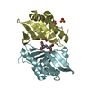
Method to determine structure: SAD / Resolution: 1.802→28.54 Å / Cor.coef. Fo:Fc: 0.959 / Cor.coef. Fo:Fc free: 0.937 / WRfactor Rfree: 0.191 / WRfactor Rwork: 0.158 / Occupancy max: 1 / Occupancy min: 0.3 / SU B: 5.291 / SU ML: 0.085 / Cross valid method: THROUGHOUT / σ(F): 0 / ESU R: 0.14 / ESU R Free: 0.131 / Stereochemistry target values: MAXIMUM LIKELIHOOD
Details: HYDROGENS HAVE BEEN ADDED IN THE RIDING POSITIONS U VALUES : WITH TLS ADDED. SAD PHASING WAS PERFORMED USING MERGED INTENSITIES FROM HKL3000 WITH THE "AUTO CORRECTIONS" OPTION FOR SCALEPACK. ...Details: HYDROGENS HAVE BEEN ADDED IN THE RIDING POSITIONS U VALUES : WITH TLS ADDED. SAD PHASING WAS PERFORMED USING MERGED INTENSITIES FROM HKL3000 WITH THE "AUTO CORRECTIONS" OPTION FOR SCALEPACK. BUCCANEER AND ARP/WARP WERE USED FOR MODEL TRACING. COOT AND THE MOLPROBITY SERVER WERE ALSO USED DURING REFINEMENT
RfactorNum. reflection% reflectionSelection details
Rfree0.2162 552 4.714 %RANDOM
Rwork0.1746 ---
obs0.177 11711 98.387 %-
Solvent computationIon probe radii: 0.8 Å / Shrinkage radii: 0.8 Å / VDW probe radii: 1.2 Å / Solvent model: MASK BULK SOLVENT
Displacement parametersBiso max: 54.08 Å2 / Biso mean: 20.391 Å2 / Biso min: 9.62 Å2
Baniso -1Baniso -2Baniso -3
1-0.149 Å20 Å20 Å2
2--0.149 Å20 Å2
3----0.298 Å2
Refinement stepCycle: LAST / Resolution: 1.802→28.54 Å
ProteinNucleic acidLigandSolventTotal
Num. atoms1057 0 17 72 1146
Refine LS restraints
Refine-IDTypeDev idealDev ideal targetNumber
X-RAY DIFFRACTIONr_bond_refined_d0.0150.0191153
X-RAY DIFFRACTIONr_bond_other_d0.0020.021095
X-RAY DIFFRACTIONr_angle_refined_deg1.4411.9421571
X-RAY DIFFRACTIONr_angle_other_deg0.87532525
X-RAY DIFFRACTIONr_dihedral_angle_1_deg4.5695143
X-RAY DIFFRACTIONr_dihedral_angle_2_deg35.81923.33357
X-RAY DIFFRACTIONr_dihedral_angle_3_deg13.90315213
X-RAY DIFFRACTIONr_dihedral_angle_4_deg18.6981510
X-RAY DIFFRACTIONr_chiral_restr0.0870.2174
X-RAY DIFFRACTIONr_gen_planes_refined0.0070.021288
X-RAY DIFFRACTIONr_gen_planes_other0.0010.02280
LS refinement shell

Refine-ID: X-RAY DIFFRACTION / Total num. of bins used: 20

Resolution (Å)Rfactor RfreeNum. reflection RfreeRfactor RworkNum. reflection RworkNum. reflection all% reflection obs (%)
1.802-1.8490.3440.23177585595.789
1.849-1.8990.287310.22277382597.455
1.899-1.9540.201430.20273579997.372
1.954-2.0140.233330.18375280297.88
2.014-2.080.254420.1669675497.878
2.08-2.1520.189320.1669073897.832
2.152-2.2330.171380.16766771698.464
2.233-2.3240.184360.16764268898.547
2.324-2.4260.215400.16762467298.81
2.426-2.5440.382250.16960864198.752
2.544-2.680.241230.16259061799.352
2.68-2.8420.154210.17655157699.306
2.842-3.0360.206270.17452755599.82
3.036-3.2760.242220.16848951499.416
3.276-3.5850.198260.16645448199.792
3.585-4.0010.188210.148423444100
4.001-4.6060.154150.14338440099.75
4.606-5.6080.247140.20933334899.713
5.608-7.7960.285110.22626828099.643
7.796-300.2280.21117718997.884
Refinement TLS params.Method: refined / Origin x: 2.6917 Å / Origin y: 32.1058 Å / Origin z: 62.9805 Å
111213212223313233
T0.0125 Å2-0.0003 Å20.0062 Å2-0.0348 Å2-0.0091 Å2--0.0064 Å2
L2.4117 °20.506 °20.2027 °2-3.0092 °20.3768 °2--1.5838 °2
S-0.0278 Å °0.1153 Å °-0.0536 Å °-0.1104 Å °0.0448 Å °-0.0278 Å °-0.0003 Å °0.0225 Å °-0.017 Å °

+
About Yorodumi

-
News

-
Feb 9, 2022. New format data for meta-information of EMDB entries

New format data for meta-information of EMDB entries

  • Version 3 of the EMDB header file is now the official format.
  • The previous official version 1.9 will be removed from the archive.

Related info.:EMDB header

External links:wwPDB to switch to version 3 of the EMDB data model

-
Aug 12, 2020. Covid-19 info

Covid-19 info

URL: https://pdbjlvh1.pdbj.org/emnavi/covid19.php

New page: Covid-19 featured information page in EM Navigator.

Related info.:Covid-19 info / Mar 5, 2020. Novel coronavirus structure data

+
Mar 5, 2020. Novel coronavirus structure data

Novel coronavirus structure data

Related info.:Yorodumi Speices / Aug 12, 2020. Covid-19 info

External links:COVID-19 featured content - PDBj / Molecule of the Month (242):Coronavirus Proteases

+
Jan 31, 2019. EMDB accession codes are about to change! (news from PDBe EMDB page)

EMDB accession codes are about to change! (news from PDBe EMDB page)

  • The allocation of 4 digits for EMDB accession codes will soon come to an end. Whilst these codes will remain in use, new EMDB accession codes will include an additional digit and will expand incrementally as the available range of codes is exhausted. The current 4-digit format prefixed with “EMD-” (i.e. EMD-XXXX) will advance to a 5-digit format (i.e. EMD-XXXXX), and so on. It is currently estimated that the 4-digit codes will be depleted around Spring 2019, at which point the 5-digit format will come into force.
  • The EM Navigator/Yorodumi systems omit the EMD- prefix.

Related info.:Q: What is EMD? / ID/Accession-code notation in Yorodumi/EM Navigator

External links:EMDB Accession Codes are Changing Soon! / Contact to PDBj

+
Jul 12, 2017. Major update of PDB

Major update of PDB

  • wwPDB released updated PDB data conforming to the new PDBx/mmCIF dictionary.
  • This is a major update changing the version number from 4 to 5, and with Remediation, in which all the entries are updated.
  • In this update, many items about electron microscopy experimental information are reorganized (e.g. em_software).
  • Now, EM Navigator and Yorodumi are based on the updated data.

External links:wwPDB Remediation / Enriched Model Files Conforming to OneDep Data Standards Now Available in the PDB FTP Archive

-
Yorodumi

Thousand views of thousand structures

  • Yorodumi is a browser for structure data from EMDB, PDB, SASBDB, etc.
  • This page is also the successor to EM Navigator detail page, and also detail information page/front-end page for Omokage search.
  • The word "yorodu" (or yorozu) is an old Japanese word meaning "ten thousand". "mi" (miru) is to see.

Related info.:EMDB / PDB / SASBDB / Comparison of 3 databanks / Yorodumi Search / Aug 31, 2016. New EM Navigator & Yorodumi / Yorodumi Papers / Jmol/JSmol / Function and homology information / Changes in new EM Navigator and Yorodumi

Read more