- PDB-4ffo: PylC in complex with phosphorylated D-ornithine -
+
Open data
ID or keywords:
Loading...
-
Basic information
Entry
Database: PDB / ID: 4ffo
Title
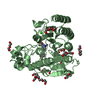
PylC in complex with phosphorylated D-ornithine
Components
PylC
Keywords
LIGASE/Reaction intermediate / amino acid / biosynthesis of pyrrolysine / isopeptide bond formation / ATP-grasp fold / Ligase / L-lysine and 3R-methyl-D-ornithine / Cytosol / LIGASE-Reaction intermediate complex
Function / homology
Function and homology information
3-methyl-D-ornithine-L-lysine ligase / pyrrolysine biosynthetic process / ligase activity / ATP binding / metal ion binding / cytosol Similarity search - Function
Resolution: 2→2.1 Å / Rmerge(I) obs: 0.471 / Mean I/σ(I) obs: 4.5 / % possible all: 99.9
-
Processing
Software
Name
Classification
XDS
datascaling
REFMAC
refinement
XDS
datareduction
XSCALE
datascaling
REFMAC
phasing
Refinement
Method to determine structure: MOLECULAR REPLACEMENT / Resolution: 2→10 Å / Cor.coef. Fo:Fc: 0.962 / Cor.coef. Fo:Fc free: 0.952 / SU B: 8.053 / SU ML: 0.1 / Cross valid method: THROUGHOUT / ESU R Free: 0.154 / Stereochemistry target values: MAXIMUM LIKELIHOOD / Details: HYDROGENS HAVE BEEN USED IF PRESENT IN THE INPUT
Rfactor
Num. reflection
% reflection
Selection details
Rfree
0.20002
1156
5 %
RANDOM
Rwork
0.15979
-
-
-
all
0.162
21964
-
-
obs
0.16187
21964
99.79 %
-
Solvent computation
Ion probe radii: 0.8 Å / Shrinkage radii: 0.8 Å / VDW probe radii: 1.2 Å / Solvent model: MASK
Displacement parameters
Biso mean: 30.712 Å2
Baniso -1
Baniso -2
Baniso -3
1-
1.11 Å2
0 Å2
0 Å2
2-
-
1.11 Å2
0 Å2
3-
-
-
-2.22 Å2
Refinement step
Cycle: LAST / Resolution: 2→10 Å
Protein
Nucleic acid
Ligand
Solvent
Total
Num. atoms
2791
0
73
260
3124
Refine LS restraints
Refine-ID
Type
Dev ideal
Dev ideal target
Number
X-RAY DIFFRACTION
r_bond_refined_d
0.007
0.02
2942
X-RAY DIFFRACTION
r_angle_refined_deg
1.33
2.017
3970
X-RAY DIFFRACTION
r_dihedral_angle_1_deg
4.998
5
350
X-RAY DIFFRACTION
r_dihedral_angle_2_deg
37.494
24.435
124
X-RAY DIFFRACTION
r_dihedral_angle_3_deg
14.231
15
505
X-RAY DIFFRACTION
r_dihedral_angle_4_deg
14.219
15
13
X-RAY DIFFRACTION
r_chiral_restr
0.073
0.2
435
X-RAY DIFFRACTION
r_gen_planes_refined
0.004
0.021
2166
X-RAY DIFFRACTION
r_rigid_bond_restr
1.353
3
2941
X-RAY DIFFRACTION
r_sphericity_free
27.369
5
91
X-RAY DIFFRACTION
r_sphericity_bonded
6.473
5
3033
LS refinement shell
Resolution: 2→2.05 Å / Total num. of bins used: 20
Rfactor
Num. reflection
% reflection
Rfree
0.289
70
-
Rwork
0.191
1375
-
obs
-
-
99.86 %
Refinement TLS params.
Method: refined / Origin x: 14.808 Å / Origin y: 4.984 Å / Origin z: 26.103 Å
11
12
13
21
22
23
31
32
33
T
0.0061 Å2
0.0016 Å2
-0.0009 Å2
-
0.004 Å2
0.0013 Å2
-
-
0.0016 Å2
L
0.255 °2
0.0424 °2
0.0049 °2
-
0.128 °2
-0.0149 °2
-
-
0.139 °2
S
-0.0114 Å °
0.0014 Å °
-0.0096 Å °
-0.0057 Å °
0.01 Å °
0.0049 Å °
0.0072 Å °
0.0117 Å °
0.0015 Å °
Refinement TLS group
ID
Refine-ID
Refine TLS-ID
Auth asym-ID
Auth seq-ID
1
X-RAY DIFFRACTION
1
A
1 - 363
2
X-RAY DIFFRACTION
1
A
901 - 905
+
About Yorodumi
-
News
-
Feb 9, 2022. New format data for meta-information of EMDB entries
New format data for meta-information of EMDB entries
Version 3 of the EMDB header file is now the official format.
The previous official version 1.9 will be removed from the archive.
In the structure databanks used in Yorodumi, some data are registered as the other names, "COVID-19 virus" and "2019-nCoV". Here are the details of the virus and the list of structure data.
Jan 31, 2019. EMDB accession codes are about to change! (news from PDBe EMDB page)
EMDB accession codes are about to change! (news from PDBe EMDB page)
The allocation of 4 digits for EMDB accession codes will soon come to an end. Whilst these codes will remain in use, new EMDB accession codes will include an additional digit and will expand incrementally as the available range of codes is exhausted. The current 4-digit format prefixed with “EMD-” (i.e. EMD-XXXX) will advance to a 5-digit format (i.e. EMD-XXXXX), and so on. It is currently estimated that the 4-digit codes will be depleted around Spring 2019, at which point the 5-digit format will come into force.
The EM Navigator/Yorodumi systems omit the EMD- prefix.
Related info.:Q: What is EMD? / ID/Accession-code notation in Yorodumi/EM Navigator
Yorodumi is a browser for structure data from EMDB, PDB, SASBDB, etc.
This page is also the successor to EM Navigator detail page, and also detail information page/front-end page for Omokage search.
The word "yorodu" (or yorozu) is an old Japanese word meaning "ten thousand". "mi" (miru) is to see.
Related info.:EMDB / PDB / SASBDB / Comparison of 3 databanks / Yorodumi Search / Aug 31, 2016. New EM Navigator & Yorodumi / Yorodumi Papers / Jmol/JSmol / Function and homology information / Changes in new EM Navigator and Yorodumi
 Movie
Movie Controller


 Open data
 Basic information
 Components
 Keywords
 Function and homology information
 Methanosarcina barkeri (archaea)
 X-RAY DIFFRACTION /
  Authors
 Citation
 Structure visualization
 Downloads & links
 Other downloads


 PDBj
PDBj


 Assembly


 Mass: 212.141 Da / Num. of mol.: 1 / Source method: obtained synthetically / Formula: C5H13N2O5P
 Mass: 427.201 Da / Num. of mol.: 1 / Source method: obtained synthetically / Formula: C10H15N5O10P2 / Comment: ADP, energy-carrying molecule*YM
 Mass: 24.305 Da / Num. of mol.: 2 / Source method: obtained synthetically / Formula: Mg
 Mass: 507.181 Da / Num. of mol.: 1 / Source method: obtained synthetically / Formula: C10H16N5O13P3 / Comment: ATP, energy-carrying molecule*YM
 Sample preparation
 / Beamline: X06SA / Wavelength: 1 Å
 Processing