 Movie
Movie Controller

[English] 日本語
 Yorodumi
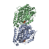
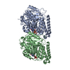
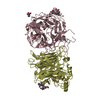
- PDB-4fdj: The molecular basis of mucopolysaccharidosis IV A, complex with GalNAc -
+ Open data

- Basic information
| Entry | Database: PDB / ID: 4fdj | |||||||||
|---|---|---|---|---|---|---|---|---|---|---|
| Title | The molecular basis of mucopolysaccharidosis IV A, complex with GalNAc | |||||||||
 Components Components | N-acetylgalactosamine-6-sulfatase | |||||||||
 Keywords Keywords | HYDROLASE / glycoprotein / enzyme replacement therapy / sulfatase / carbohydrate-binding protein / formylglycine / N-linked glycosylation / lysosomal enzyme | |||||||||
| Function / homology |  Function and homology information Function and homology informationN-acetylgalactosamine-6-sulfatase / N-acetylgalactosamine-6-sulfatase activity / MPS IV - Morquio syndrome A / N-acetylgalactosamine-4-sulfatase activity / chondroitin sulfate proteoglycan catabolic process / Keratan sulfate degradation / sulfuric ester hydrolase activity / arylsulfatase activity / lysosomal lumen / azurophil granule lumen ...N-acetylgalactosamine-6-sulfatase / N-acetylgalactosamine-6-sulfatase activity / MPS IV - Morquio syndrome A / N-acetylgalactosamine-4-sulfatase activity / chondroitin sulfate proteoglycan catabolic process / Keratan sulfate degradation / sulfuric ester hydrolase activity / arylsulfatase activity / lysosomal lumen / azurophil granule lumen / Neutrophil degranulation / extracellular exosome / extracellular region / metal ion binding Similarity search - Function | |||||||||
| Biological species |  Homo sapiens (human) Homo sapiens (human) | |||||||||
| Method |  X-RAY DIFFRACTION / SYNCHROTRON / FOURIER SYNTHESIS / Resolution: 2.81 Å X-RAY DIFFRACTION / SYNCHROTRON / FOURIER SYNTHESIS / Resolution: 2.81 Å | |||||||||
 Authors Authors | Rivera-Colon, Y. / Garman, S.C. | |||||||||
 Citation Citation | Journal: J.Mol.Biol. / Year: 2012 Title: The Structure of Human GALNS Reveals the Molecular Basis for Mucopolysaccharidosis IV A. Authors: Rivera-Colon, Y. / Schutsky, E.K. / Kita, A.Z. / Garman, S.C. | |||||||||
| History |
|
- Structure visualization
| Structure viewer | Molecule: MolmilJmol/JSmol |
|---|
- Downloads & links
-Download
| PDBx/mmCIF format | 4fdj.cif.gz | 405.7 KB | Display | PDBx/mmCIF format |
|---|---|---|---|---|
| PDB format | pdb4fdj.ent.gz | 331.9 KB | Display | PDB format |
| PDBx/mmJSON format | 4fdj.json.gz | Tree view | PDBx/mmJSON format | |
| Others |  Other downloads Other downloads |
-Validation report
| Arichive directory | https://data.pdbj.org/pub/pdb/validation_reports/fd/4fdjftp://data.pdbj.org/pub/pdb/validation_reports/fd/4fdj | HTTPS FTP |
|---|
-Related structure data
| Related structure data | 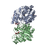 4fdiSC S: Starting model for refinement C: citing same article ( |
|---|---|
| Similar structure data |


-Links
 PDBj
PDBj
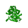
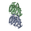
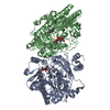
- Assembly
| Deposited unit | 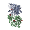
| |||||||||||||||||||||||||||||||||||||||||||||||||||||||||||||||||||||||||||||||||||||||
|---|---|---|---|---|---|---|---|---|---|---|---|---|---|---|---|---|---|---|---|---|---|---|---|---|---|---|---|---|---|---|---|---|---|---|---|---|---|---|---|---|---|---|---|---|---|---|---|---|---|---|---|---|---|---|---|---|---|---|---|---|---|---|---|---|---|---|---|---|---|---|---|---|---|---|---|---|---|---|---|---|---|---|---|---|---|---|---|---|
| 1 |
| |||||||||||||||||||||||||||||||||||||||||||||||||||||||||||||||||||||||||||||||||||||||
| Unit cell |
| |||||||||||||||||||||||||||||||||||||||||||||||||||||||||||||||||||||||||||||||||||||||
| Noncrystallographic symmetry (NCS) | NCS domain:
NCS domain segments:
|
-Components
| #1: Protein | Mass: 56301.426 Da / Num. of mol.: 2 / Fragment: UNP residues 27-522 Source method: isolated from a genetically manipulated source Details: Selected with blastocidin / Source: (gene. exp.) Homo sapiens (human) / Gene: GALNS / Plasmid: pIB/V5-His-TOPO TA / Production host:  Trichoplusia ni (cabbage looper) / Strain (production host): HI-FIVE Trichoplusia ni (cabbage looper) / Strain (production host): HI-FIVEReferences: UniProt: P34059, N-acetylgalactosamine-6-sulfatase #2: Sugar | ChemComp-NAG /   Type: D-saccharide, beta linking / Mass: 221.208 Da / Num. of mol.: 4 Type: D-saccharide, beta linking / Mass: 221.208 Da / Num. of mol.: 4Source method: isolated from a genetically manipulated source Formula: C8H15NO6 #3: Chemical |   Mass: 40.078 Da / Num. of mol.: 2 / Source method: obtained synthetically / Formula: Ca Mass: 40.078 Da / Num. of mol.: 2 / Source method: obtained synthetically / Formula: Ca#4: Sugar | 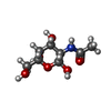  Type: D-saccharide, beta linking / Mass: 221.208 Da / Num. of mol.: 2 Type: D-saccharide, beta linking / Mass: 221.208 Da / Num. of mol.: 2Source method: isolated from a genetically manipulated source Formula: C8H15NO6 #5: Water | ChemComp-HOH / |  Mass: 18.015 Da / Num. of mol.: 205 / Source method: isolated from a natural source / Formula: H2O Mass: 18.015 Da / Num. of mol.: 205 / Source method: isolated from a natural source / Formula: H2OHas protein modification | Y | |
|---|
-Experimental details
-Experiment
| Experiment | Method: X-RAY DIFFRACTION / Number of used crystals: 1 |
|---|
- Sample preparation
| Crystal | Density Matthews: 2.39 Å3/Da / Density % sol: 48.49 % |
|---|---|
| Crystal grow | Temperature: 293 K / Method: vapor diffusion, hanging drop / pH: 5 Details: 10-15% PEG6000, 0.1 M citric acid, pH 5.0, VAPOR DIFFUSION, HANGING DROP, temperature 293K |
-Data collection
| Diffraction | Mean temperature: 100 K |
|---|---|
| Diffraction source | Source: SYNCHROTRON / Site: NSLS  / Beamline: X6A / Wavelength: 1.1 Å / Beamline: X6A / Wavelength: 1.1 Å |
| Detector | Type: ADSC QUANTUM 270 / Detector: CCD / Date: Aug 23, 2011 / Details: toroidal focusing mirror |
| Radiation | Monochromator: Si(111) channel cut / Protocol: SINGLE WAVELENGTH / Monochromatic (M) / Laue (L): M / Scattering type: x-ray |
| Radiation wavelength | Wavelength: 1.1 Å / Relative weight: 1 |
| Reflection | Resolution: 2.8→50 Å / Num. obs: 24690 / % possible obs: 96.4 % / Redundancy: 2.9 % / Biso Wilson estimate: 39.07 Å2 / Rsym value: 0.143 / Χ2: 1.202 / Net I/σ(I): 8.6 |
| Reflection shell | Resolution: 2.8→2.85 Å / Redundancy: 2.4 % / Rmerge(I) obs: 0.278 / Num. unique all: 1055 / Χ2: 0.806 / % possible all: 83.7 |
- Processing
| Software |
| |||||||||||||||||||||||||||||||||||||||||||||||||||||||||||||||||||||||||||||||||||||||||||||||||||||||||||||||||||||||||||||||||||||||||||||||||||||||||||||||||||||||||||||||
|---|---|---|---|---|---|---|---|---|---|---|---|---|---|---|---|---|---|---|---|---|---|---|---|---|---|---|---|---|---|---|---|---|---|---|---|---|---|---|---|---|---|---|---|---|---|---|---|---|---|---|---|---|---|---|---|---|---|---|---|---|---|---|---|---|---|---|---|---|---|---|---|---|---|---|---|---|---|---|---|---|---|---|---|---|---|---|---|---|---|---|---|---|---|---|---|---|---|---|---|---|---|---|---|---|---|---|---|---|---|---|---|---|---|---|---|---|---|---|---|---|---|---|---|---|---|---|---|---|---|---|---|---|---|---|---|---|---|---|---|---|---|---|---|---|---|---|---|---|---|---|---|---|---|---|---|---|---|---|---|---|---|---|---|---|---|---|---|---|---|---|---|---|---|---|---|---|
| Refinement | Method to determine structure: FOURIER SYNTHESIS Starting model: PDB ENTRY 4FDI Resolution: 2.81→45.31 Å / Cor.coef. Fo:Fc: 0.918 / Cor.coef. Fo:Fc free: 0.868 / Occupancy max: 1 / Occupancy min: 1 / SU B: 39.286 / SU ML: 0.34 / Cross valid method: THROUGHOUT / σ(F): 0 / ESU R Free: 0.427 / Stereochemistry target values: MAXIMUM LIKELIHOOD Details: HYDROGENS HAVE BEEN ADDED IN THE RIDING POSITIONS U VALUES : WITH TLS ADDED
| |||||||||||||||||||||||||||||||||||||||||||||||||||||||||||||||||||||||||||||||||||||||||||||||||||||||||||||||||||||||||||||||||||||||||||||||||||||||||||||||||||||||||||||||
| Solvent computation | Ion probe radii: 0.8 Å / Shrinkage radii: 0.8 Å / VDW probe radii: 1.4 Å / Solvent model: MASK | |||||||||||||||||||||||||||||||||||||||||||||||||||||||||||||||||||||||||||||||||||||||||||||||||||||||||||||||||||||||||||||||||||||||||||||||||||||||||||||||||||||||||||||||
| Displacement parameters | Biso max: 110.7 Å2 / Biso mean: 39.9954 Å2 / Biso min: 10.97 Å2
| |||||||||||||||||||||||||||||||||||||||||||||||||||||||||||||||||||||||||||||||||||||||||||||||||||||||||||||||||||||||||||||||||||||||||||||||||||||||||||||||||||||||||||||||
| Refinement step | Cycle: LAST / Resolution: 2.81→45.31 Å
| |||||||||||||||||||||||||||||||||||||||||||||||||||||||||||||||||||||||||||||||||||||||||||||||||||||||||||||||||||||||||||||||||||||||||||||||||||||||||||||||||||||||||||||||
| Refine LS restraints |
| |||||||||||||||||||||||||||||||||||||||||||||||||||||||||||||||||||||||||||||||||||||||||||||||||||||||||||||||||||||||||||||||||||||||||||||||||||||||||||||||||||||||||||||||
| Refine LS restraints NCS | Dom-ID: 1 / Auth asym-ID: A / Ens-ID: 1 / Refine-ID: X-RAY DIFFRACTION
| |||||||||||||||||||||||||||||||||||||||||||||||||||||||||||||||||||||||||||||||||||||||||||||||||||||||||||||||||||||||||||||||||||||||||||||||||||||||||||||||||||||||||||||||
| LS refinement shell | Resolution: 2.81→2.879 Å / Total num. of bins used: 20
| |||||||||||||||||||||||||||||||||||||||||||||||||||||||||||||||||||||||||||||||||||||||||||||||||||||||||||||||||||||||||||||||||||||||||||||||||||||||||||||||||||||||||||||||
| Refinement TLS params. | Method: refined / Refine-ID: X-RAY DIFFRACTION
| |||||||||||||||||||||||||||||||||||||||||||||||||||||||||||||||||||||||||||||||||||||||||||||||||||||||||||||||||||||||||||||||||||||||||||||||||||||||||||||||||||||||||||||||
| Refinement TLS group |
|
