 Movie
Movie Controller

[English] 日本語
 Yorodumi
- PDB-4fd0: Crystal structure of a putative cell surface protein (BACCAC_0370... -
+ Open data

- Basic information
| Entry | Database: PDB / ID: 4fd0 | ||||||
|---|---|---|---|---|---|---|---|
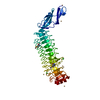
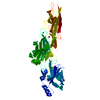
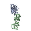
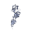
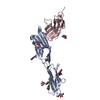
| Title | Crystal structure of a putative cell surface protein (BACCAC_03700) from Bacteroides caccae ATCC 43185 at 2.07 A resolution | ||||||
 Components Components | Leucine rich hypothetical protein | ||||||
 Keywords Keywords | STRUCTURAL GENOMICS / UNKNOWN FUNCTION / putative cell surface protein / Big3 domain / LRR domain / Joint Center for Structural Genomics / JCSG / Protein Structure Initiative / PSI-BIOLOGY | ||||||
| Function / homology |  Function and homology information Function and homology informationImmunoglobulin-like - #3630 / Bacterial Ig-like domain (group 3) / Ig-like domain, bacterial type / BspA type Leucine rich repeat region / BspA type Leucine rich repeat region (6 copies) / Leucine-rich repeat, LRR (right-handed beta-alpha superhelix) / Ribonuclease Inhibitor / Alpha-Beta Horseshoe / Leucine-rich repeat domain superfamily / Immunoglobulin-like ...Immunoglobulin-like - #3630 / Bacterial Ig-like domain (group 3) / Ig-like domain, bacterial type / BspA type Leucine rich repeat region / BspA type Leucine rich repeat region (6 copies) / Leucine-rich repeat, LRR (right-handed beta-alpha superhelix) / Ribonuclease Inhibitor / Alpha-Beta Horseshoe / Leucine-rich repeat domain superfamily / Immunoglobulin-like / Sandwich / Mainly Beta / Alpha Beta Similarity search - Domain/homology | ||||||
| Biological species |  Bacteroides caccae (bacteria) Bacteroides caccae (bacteria) | ||||||
| Method |  X-RAY DIFFRACTION / SYNCHROTRON / MAD / Resolution: 2.07 Å X-RAY DIFFRACTION / SYNCHROTRON / MAD / Resolution: 2.07 Å | ||||||
 Authors Authors | Joint Center for Structural Genomics (JCSG) | ||||||
 Citation Citation | Journal: To be published Title: Crystal structure of a leucine rich hypothetical protein (BACCAC_03700) from Bacteroides caccae ATCC 43185 at 2.07 A resolution (CASP Target) Authors: Joint Center for Structural Genomics (JCSG) | ||||||
| History |
|
- Structure visualization
| Structure viewer | Molecule: MolmilJmol/JSmol |
|---|
- Downloads & links
-Download
| PDBx/mmCIF format | 4fd0.cif.gz | 105.4 KB | Display | PDBx/mmCIF format |
|---|---|---|---|---|
| PDB format | pdb4fd0.ent.gz | 77.8 KB | Display | PDB format |
| PDBx/mmJSON format | 4fd0.json.gz | Tree view | PDBx/mmJSON format | |
| Others |  Other downloads Other downloads |
-Validation report
| Arichive directory | https://data.pdbj.org/pub/pdb/validation_reports/fd/4fd0ftp://data.pdbj.org/pub/pdb/validation_reports/fd/4fd0 | HTTPS FTP |
|---|
-Related structure data
| Related structure data | |
|---|---|
| Similar structure data | |
| Other databases |


-Links
 PDBj
PDBj- Assembly
| Deposited unit | 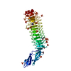
| ||||||||
|---|---|---|---|---|---|---|---|---|---|
| 1 |
| ||||||||
| Unit cell |
|
-Components
| #1: Protein | Mass: 44710.203 Da / Num. of mol.: 1 / Fragment: UNP residues 26-428 Source method: isolated from a genetically manipulated source Source: (gene. exp.) Bacteroides caccae (bacteria) / Gene: BACCAC_03700 / Plasmid: SpeedET / Production host: | ||||||||
|---|---|---|---|---|---|---|---|---|---|
| #2: Chemical | ChemComp-SO4 /   Mass: 96.063 Da / Num. of mol.: 16 / Source method: obtained synthetically / Formula: SO4 Mass: 96.063 Da / Num. of mol.: 16 / Source method: obtained synthetically / Formula: SO4#3: Chemical | ChemComp-CL / |   Mass: 35.453 Da / Num. of mol.: 1 / Source method: obtained synthetically / Formula: Cl Mass: 35.453 Da / Num. of mol.: 1 / Source method: obtained synthetically / Formula: Cl#4: Water | ChemComp-HOH / |  Mass: 18.015 Da / Num. of mol.: 460 / Source method: isolated from a natural source / Formula: H2O Mass: 18.015 Da / Num. of mol.: 460 / Source method: isolated from a natural source / Formula: H2OHas protein modification | Y | Sequence details | THE CONSTRUCT (RESIDUES 26-428) WAS EXPRESSED WITH A PURIFICATION TAG MGSDKIHHHHHHENLYFQG. THE TAG ...THE CONSTRUCT (RESIDUES 26-428) WAS EXPRESSED WITH A PURIFICATI | |
-Experimental details
-Experiment
| Experiment | Method: X-RAY DIFFRACTION / Number of used crystals: 1 |
|---|
- Sample preparation
| Crystal | Density Matthews: 3.91 Å3/Da / Density % sol: 68.53 % |
|---|---|
| Crystal grow | Temperature: 277 K / Method: vapor diffusion, sitting drop / pH: 10.5 Details: 0.200M lithium sulfate, 2.00M ammonium sulfate, 0.1M CAPS pH 10.5, NANODROP, VAPOR DIFFUSION, SITTING DROP, temperature 277K |
-Data collection
| Diffraction | Mean temperature: 100 K | |||||||||||||||||||||||||||||||||||||||||||||||||||||||||||||||||||||||||||||
|---|---|---|---|---|---|---|---|---|---|---|---|---|---|---|---|---|---|---|---|---|---|---|---|---|---|---|---|---|---|---|---|---|---|---|---|---|---|---|---|---|---|---|---|---|---|---|---|---|---|---|---|---|---|---|---|---|---|---|---|---|---|---|---|---|---|---|---|---|---|---|---|---|---|---|---|---|---|---|
| Diffraction source | Source: SYNCHROTRON / Site: SSRL  / Beamline: BL11-1 / Wavelength: 0.97932,0.91837,0.97862 / Beamline: BL11-1 / Wavelength: 0.97932,0.91837,0.97862 | |||||||||||||||||||||||||||||||||||||||||||||||||||||||||||||||||||||||||||||
| Detector | Type: DECTRIS PILATUS 6M / Detector: PIXEL / Date: Apr 4, 2012 Details: Flat mirror (vertical focusing); single crystal Si(111) bent monochromator (horizontal focusing) | |||||||||||||||||||||||||||||||||||||||||||||||||||||||||||||||||||||||||||||
| Radiation | Monochromator: single crystal Si(111) bent / Protocol: MAD / Monochromatic (M) / Laue (L): M / Scattering type: x-ray | |||||||||||||||||||||||||||||||||||||||||||||||||||||||||||||||||||||||||||||
| Radiation wavelength |
| |||||||||||||||||||||||||||||||||||||||||||||||||||||||||||||||||||||||||||||
| Reflection | Resolution: 2.07→46.029 Å / Num. obs: 42010 / % possible obs: 97.4 % / Observed criterion σ(I): -3 / Biso Wilson estimate: 32.803 Å2 / Rmerge(I) obs: 0.079 / Net I/σ(I): 12.27 | |||||||||||||||||||||||||||||||||||||||||||||||||||||||||||||||||||||||||||||
| Reflection shell |
|
-Phasing
| Phasing | Method: MAD |
|---|
- Processing
| Software |
| ||||||||||||||||||||||||||||||||||||||||||||||||||||||||||||||||||||||||||||||||||||||||||||||||||||||||||||
|---|---|---|---|---|---|---|---|---|---|---|---|---|---|---|---|---|---|---|---|---|---|---|---|---|---|---|---|---|---|---|---|---|---|---|---|---|---|---|---|---|---|---|---|---|---|---|---|---|---|---|---|---|---|---|---|---|---|---|---|---|---|---|---|---|---|---|---|---|---|---|---|---|---|---|---|---|---|---|---|---|---|---|---|---|---|---|---|---|---|---|---|---|---|---|---|---|---|---|---|---|---|---|---|---|---|---|---|---|---|
| Refinement | Method to determine structure: MAD / Resolution: 2.07→46.029 Å / Cor.coef. Fo:Fc: 0.9434 / Cor.coef. Fo:Fc free: 0.9319 / Occupancy max: 1 / Occupancy min: 0.3 / Cross valid method: THROUGHOUT / σ(F): 0 Details: 1. A MET-INHIBITION PROTOCOL WAS USED FOR SELENOMETHIONINE INCORPORATION DURING PROTEIN EXPRESSION. THE OCCUPANCY OF THE SE ATOMS IN THE MSE RESIDUES WAS REDUCED TO 0.75 FOR THE REDUCED ...Details: 1. A MET-INHIBITION PROTOCOL WAS USED FOR SELENOMETHIONINE INCORPORATION DURING PROTEIN EXPRESSION. THE OCCUPANCY OF THE SE ATOMS IN THE MSE RESIDUES WAS REDUCED TO 0.75 FOR THE REDUCED SCATTERING POWER DUE TO PARTIAL S-MET INCORPORATION. 2. THE MAD PHASES WERE USED AS RESTRAINTS DURING REFINEMENT. 3. CHLORIDE, SULFATE MODELED WERE PRESENT IN THE PROTEIN/CRYSTALLIZATION CONDITIONS.
| ||||||||||||||||||||||||||||||||||||||||||||||||||||||||||||||||||||||||||||||||||||||||||||||||||||||||||||
| Displacement parameters | Biso max: 100.83 Å2 / Biso mean: 37.2084 Å2 / Biso min: 19.22 Å2
| ||||||||||||||||||||||||||||||||||||||||||||||||||||||||||||||||||||||||||||||||||||||||||||||||||||||||||||
| Refine analyze | Luzzati coordinate error obs: 0.231 Å | ||||||||||||||||||||||||||||||||||||||||||||||||||||||||||||||||||||||||||||||||||||||||||||||||||||||||||||
| Refinement step | Cycle: LAST / Resolution: 2.07→46.029 Å
| ||||||||||||||||||||||||||||||||||||||||||||||||||||||||||||||||||||||||||||||||||||||||||||||||||||||||||||
| Refine LS restraints |
| ||||||||||||||||||||||||||||||||||||||||||||||||||||||||||||||||||||||||||||||||||||||||||||||||||||||||||||
| LS refinement shell | Resolution: 2.07→2.12 Å / Total num. of bins used: 20
|
