 Movie
Movie Controller

[English] 日本語
 Yorodumi
- PDB-4fbv: Crystal structure of the Myxococcus Xanthus hemagglutinin in comp... -
+ Open data

- Basic information
| Entry | Database: PDB / ID: 4fbv | |||||||||
|---|---|---|---|---|---|---|---|---|---|---|
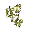
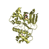
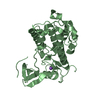
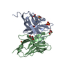
| Title | Crystal structure of the Myxococcus Xanthus hemagglutinin in complex with a3,a6-mannopentaose | |||||||||
 Components Components | Myxobacterial hemagglutinin | |||||||||
 Keywords Keywords | Carbohydrate binding protein / beta-barrel / HIV-inactivating | |||||||||
| Function / homology |  Function and homology information Function and homology information | |||||||||
| Biological species |  Myxococcus xanthus (bacteria) Myxococcus xanthus (bacteria) | |||||||||
| Method |  X-RAY DIFFRACTION / MOLECULAR REPLACEMENT / Resolution: 1.76 Å X-RAY DIFFRACTION / MOLECULAR REPLACEMENT / Resolution: 1.76 Å | |||||||||
 Authors Authors | Koharudin, L.M.I. / Gronenborn, A.M. | |||||||||
 Citation Citation | Journal: J.Biol.Chem. / Year: 2012 Title: Structural Insights into the Anti-HIV Activity of the Oscillatoria agardhii Agglutinin Homolog Lectin Family. Authors: Koharudin, L.M. / Kollipara, S. / Aiken, C. / Gronenborn, A.M. #1: Journal: Structure / Year: 2011Title: Structural basis of the anti-HIV activity of the cyanobacterial Oscillatoria Agardhii agglutinin. Authors: Koharudin, L.M. / Gronenborn, A.M. | |||||||||
| History |
|
- Structure visualization
| Structure viewer | Molecule: MolmilJmol/JSmol |
|---|
- Downloads & links
-Download
| PDBx/mmCIF format | 4fbv.cif.gz | 74.2 KB | Display | PDBx/mmCIF format |
|---|---|---|---|---|
| PDB format | pdb4fbv.ent.gz | 54.8 KB | Display | PDB format |
| PDBx/mmJSON format | 4fbv.json.gz | Tree view | PDBx/mmJSON format | |
| Others |  Other downloads Other downloads |
-Validation report
| Arichive directory | https://data.pdbj.org/pub/pdb/validation_reports/fb/4fbvftp://data.pdbj.org/pub/pdb/validation_reports/fb/4fbv | HTTPS FTP |
|---|
-Related structure data
| Related structure data |  4fboC  4fbrSC S: Starting model for refinement C: citing same article ( |
|---|---|
| Similar structure data |


-Links
 PDBj
PDBj


- Assembly
| Deposited unit | 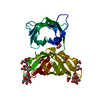
| ||||||||
|---|---|---|---|---|---|---|---|---|---|
| 1 |
| ||||||||
| Unit cell |
|
-Components
-Protein , 1 types, 1 molecules A
| #1: Protein | Mass: 27936.654 Da / Num. of mol.: 1 Source method: isolated from a genetically manipulated source Source: (gene. exp.) Myxococcus xanthus (bacteria) / Gene: mbhA / Plasmid: pET26b(+) / Production host: |
|---|
-Sugars , 3 types, 3 molecules 
| #2: Polysaccharide | alpha-D-mannopyranose-(1-3)-[alpha-D-mannopyranose-(1-6)]alpha-D-mannopyranose-(1-6)-beta-D-mannopyranose Source method: isolated from a genetically manipulated source |
|---|---|
| #3: Polysaccharide | alpha-D-mannopyranose-(1-3)-[alpha-D-mannopyranose-(1-6)]alpha-D-mannopyranose-(1-6)-[alpha-D- ...alpha-D-mannopyranose-(1-3)-[alpha-D-mannopyranose-(1-6)]alpha-D-mannopyranose-(1-6)-[alpha-D-mannopyranose-(1-3)]beta-D-mannopyranose Source method: isolated from a genetically manipulated source |
| #4: Sugar | ChemComp-MAN /  Type: D-saccharide, alpha linking / Mass: 180.156 Da / Num. of mol.: 1 Type: D-saccharide, alpha linking / Mass: 180.156 Da / Num. of mol.: 1Source method: isolated from a genetically manipulated source Formula: C6H12O6 |
-Non-polymers , 2 types, 201 molecules 

| #5: Chemical | ChemComp-EDO /  Mass: 62.068 Da / Num. of mol.: 4 / Source method: obtained synthetically / Formula: C2H6O2 Mass: 62.068 Da / Num. of mol.: 4 / Source method: obtained synthetically / Formula: C2H6O2#6: Water | ChemComp-HOH / | Mass: 18.015 Da / Num. of mol.: 197 / Source method: isolated from a natural source / Formula: H2O |
|---|
-Experimental details
-Experiment
| Experiment | Method: X-RAY DIFFRACTION / Number of used crystals: 1 |
|---|
- Sample preparation
| Crystal | Density Matthews: 2.48 Å3/Da / Density % sol: 50.37 % |
|---|---|
| Crystal grow | Temperature: 298 K / Method: vapor diffusion, sitting drop / pH: 8 Details: 0.05 M monobasic potassium phosphate and 20% w/v polyethylene glycol 3350, pH 8, VAPOR DIFFUSION, SITTING DROP, temperature 298K |
-Data collection
| Diffraction | Mean temperature: 93 K |
|---|---|
| Diffraction source | Source: ROTATING ANODE / Type: RIGAKU FR-E SUPERBRIGHT / Wavelength: 1.5418 Å |
| Detector | Type: RIGAKU SATURN 944 / Detector: CCD / Date: Jul 26, 2011 |
| Radiation | Monochromator: MIRRORS / Protocol: SINGLE WAVELENGTH / Monochromatic (M) / Laue (L): M / Scattering type: x-ray |
| Radiation wavelength | Wavelength: 1.5418 Å / Relative weight: 1 |
| Reflection | Resolution: 1.76→33.82 Å / Num. all: 28299 / Num. obs: 27394 / % possible obs: 96.8 % / Observed criterion σ(F): 1 / Observed criterion σ(I): 3.8 / Redundancy: 6.63 % / Rmerge(I) obs: 0.053 / Net I/σ(I): 22.1 |
| Reflection shell | Resolution: 1.76→1.82 Å / Redundancy: 2.53 % / Rmerge(I) obs: 0.206 / Mean I/σ(I) obs: 3.8 / Num. unique all: 2789 / % possible all: 81.5 |
- Processing
| Software |
| |||||||||||||||||||||||||||||||||||||||||||||||||||||||||||||||||
|---|---|---|---|---|---|---|---|---|---|---|---|---|---|---|---|---|---|---|---|---|---|---|---|---|---|---|---|---|---|---|---|---|---|---|---|---|---|---|---|---|---|---|---|---|---|---|---|---|---|---|---|---|---|---|---|---|---|---|---|---|---|---|---|---|---|---|
| Refinement | Method to determine structure: MOLECULAR REPLACEMENT Starting model: PDB entry 4FBR Resolution: 1.76→33.82 Å / Cor.coef. Fo:Fc: 0.958 / Cor.coef. Fo:Fc free: 0.949 / SU B: 2.388 / SU ML: 0.077 / Cross valid method: THROUGHOUT / σ(I): 3.8 / ESU R: 0.126 / ESU R Free: 0.116 / Stereochemistry target values: MAXIMUM LIKELIHOOD / Details: HYDROGENS HAVE BEEN ADDED IN THE RIDING POSITIONS
| |||||||||||||||||||||||||||||||||||||||||||||||||||||||||||||||||
| Solvent computation | Ion probe radii: 0.8 Å / Shrinkage radii: 0.8 Å / VDW probe radii: 1.4 Å / Solvent model: BABINET MODEL WITH MASK | |||||||||||||||||||||||||||||||||||||||||||||||||||||||||||||||||
| Displacement parameters | Biso mean: 24.264 Å2
| |||||||||||||||||||||||||||||||||||||||||||||||||||||||||||||||||
| Refinement step | Cycle: LAST / Resolution: 1.76→33.82 Å
| |||||||||||||||||||||||||||||||||||||||||||||||||||||||||||||||||
| Refine LS restraints |
| |||||||||||||||||||||||||||||||||||||||||||||||||||||||||||||||||
| LS refinement shell | Resolution: 1.76→1.806 Å / Total num. of bins used: 20
|
