 Movie
Movie Controller

[English] 日本語
 Yorodumi
- PDB-4f86: Structure analysis of Geranyl diphosphate methyltransferase in co... -
+ Open data

- Basic information
| Entry | Database: PDB / ID: 4f86 | ||||||
|---|---|---|---|---|---|---|---|
| Title | Structure analysis of Geranyl diphosphate methyltransferase in complex with GPP and sinefungin | ||||||
 Components Components | Geranyl diphosphate 2-C-methyltransferase | ||||||
 Keywords Keywords | TRANSFERASE / alpha/beta / Rossmann Fold / methyltransferase | ||||||
| Function / homology |  Function and homology information Function and homology informationgeranyl diphosphate 2-C-methyltransferase / C-methyltransferase activity / terpene metabolic process / S-adenosylmethionine-dependent methyltransferase activity / S-adenosyl-L-methionine binding / methylation / magnesium ion binding Similarity search - Function | ||||||
| Biological species |  Streptomyces lasaliensis (bacteria) Streptomyces lasaliensis (bacteria) | ||||||
| Method |  X-RAY DIFFRACTION / SYNCHROTRON / MOLECULAR REPLACEMENT / Resolution: 3 Å X-RAY DIFFRACTION / SYNCHROTRON / MOLECULAR REPLACEMENT / Resolution: 3 Å | ||||||
 Authors Authors | Ariyawutthiphan, O. / Ose, T. / Minami, A. / Gao, Y.G. / Yao, M. / Oikawa, H. / Tanaka, I. | ||||||
 Citation Citation | Journal: Acta Crystallogr.,Sect.D / Year: 2012 Title: Structure analysis of geranyl pyrophosphate methyltransferase and the proposed reaction mechanism of SAM-dependent C-methylation Authors: Ariyawutthiphan, O. / Ose, T. / Minami, A. / Sinde, S. / Tsuda, M. / Gao, Y.-G. / Yao, M. / Oikawa, H. / Tanaka, I. | ||||||
| History |
|
- Structure visualization
| Structure viewer | Molecule: MolmilJmol/JSmol |
|---|
- Downloads & links
-Download
| PDBx/mmCIF format | 4f86.cif.gz | 636.7 KB | Display | PDBx/mmCIF format |
|---|---|---|---|---|
| PDB format | pdb4f86.ent.gz | 522.8 KB | Display | PDB format |
| PDBx/mmJSON format | 4f86.json.gz | Tree view | PDBx/mmJSON format | |
| Others |  Other downloads Other downloads |
-Validation report
| Arichive directory | https://data.pdbj.org/pub/pdb/validation_reports/f8/4f86ftp://data.pdbj.org/pub/pdb/validation_reports/f8/4f86 | HTTPS FTP |
|---|
-Related structure data
| Related structure data |  4f84C  4f85C 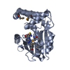 3busS C: citing same article ( S: Starting model for refinement |
|---|---|
| Similar structure data |

-Links
 PDBj
PDBj
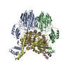
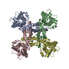
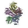
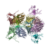
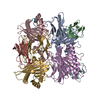
- Assembly
| Deposited unit | 
| ||||||||
|---|---|---|---|---|---|---|---|---|---|
| 1 | 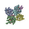
| ||||||||
| 2 | 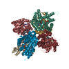
| ||||||||
| Unit cell |
|
-Components
| #1: Protein | Mass: 35092.070 Da / Num. of mol.: 12 Source method: isolated from a genetically manipulated source Source: (gene. exp.) Streptomyces lasaliensis (bacteria) / Gene: gdpmt / Plasmid: pET26b / Production host: References: UniProt: D3KYU3, Transferases; Transferring one-carbon groups; Methyltransferases #2: Chemical | ChemComp-SFG /   Mass: 381.387 Da / Num. of mol.: 12 / Source method: obtained synthetically / Formula: C15H23N7O5 Mass: 381.387 Da / Num. of mol.: 12 / Source method: obtained synthetically / Formula: C15H23N7O5#3: Chemical | ChemComp-GPP / 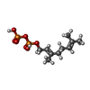  Mass: 314.209 Da / Num. of mol.: 12 / Source method: obtained synthetically / Formula: C10H20O7P2 Mass: 314.209 Da / Num. of mol.: 12 / Source method: obtained synthetically / Formula: C10H20O7P2#4: Chemical | ChemComp-MG /   Mass: 24.305 Da / Num. of mol.: 12 / Source method: obtained synthetically / Formula: Mg Mass: 24.305 Da / Num. of mol.: 12 / Source method: obtained synthetically / Formula: Mg |
|---|
-Experimental details
-Experiment
| Experiment | Method: X-RAY DIFFRACTION / Number of used crystals: 1 |
|---|
- Sample preparation
| Crystal | Density Matthews: 2.58 Å3/Da / Density % sol: 52.24 % |
|---|---|
| Crystal grow | Temperature: 293 K / Method: vapor diffusion, hanging drop / pH: 6.7 Details: 1.3M lithium sulfate, 1mM sperminero, 30mM magnesium chloride, 50mM sodium cacodylate, pH 6.7, VAPOR DIFFUSION, HANGING DROP, temperature 293K |
-Data collection
| Diffraction | Mean temperature: 100 K |
|---|---|
| Diffraction source | Source: SYNCHROTRON / Site: SPring-8  / Beamline: BL41XU / Wavelength: 1 Å / Beamline: BL41XU / Wavelength: 1 Å |
| Detector | Type: ADSC QUANTUM 315r / Detector: CCD / Date: Nov 21, 2009 / Details: mirror |
| Radiation | Monochromator: silicon / Protocol: SINGLE WAVELENGTH / Monochromatic (M) / Laue (L): M / Scattering type: x-ray |
| Radiation wavelength | Wavelength: 1 Å / Relative weight: 1 |
| Reflection | Resolution: 3→50 Å / Num. obs: 82669 / % possible obs: 96.3 % / Observed criterion σ(I): 0 / Redundancy: 3.5 % / Biso Wilson estimate: 39.5 Å2 / Rmerge(I) obs: 0.118 |
| Reflection shell | Resolution: 3→3.05 Å / Redundancy: 3.5 % / Rmerge(I) obs: 0.359 / Mean I/σ(I) obs: 1.7 / Num. unique all: 4153 / % possible all: 95.8 |
- Processing
| Software |
| ||||||||||||||||||||||||||||||||
|---|---|---|---|---|---|---|---|---|---|---|---|---|---|---|---|---|---|---|---|---|---|---|---|---|---|---|---|---|---|---|---|---|---|
| Refinement | Method to determine structure: MOLECULAR REPLACEMENT Starting model: 3bus Resolution: 3→46.51 Å / Rfactor Rfree error: 0.005 / Occupancy max: 1 / Occupancy min: 1 / Data cutoff high absF: 2735189 / Data cutoff low absF: 0 / Isotropic thermal model: OVERALL / Cross valid method: THROUGHOUT / σ(F): 0 / Stereochemistry target values: Engh & Huber / Details: BULK SOLVENT MODEL USED
| ||||||||||||||||||||||||||||||||
| Solvent computation | Solvent model: FLAT MODEL / Bsol: -6.8095 Å2 / ksol: 0.25 e/Å3 | ||||||||||||||||||||||||||||||||
| Displacement parameters | Biso max: 98.47 Å2 / Biso mean: 50.5488 Å2 / Biso min: 9.97 Å2
| ||||||||||||||||||||||||||||||||
| Refine analyze |
| ||||||||||||||||||||||||||||||||
| Refinement step | Cycle: LAST / Resolution: 3→46.51 Å
| ||||||||||||||||||||||||||||||||
| Refine LS restraints |
| ||||||||||||||||||||||||||||||||
| LS refinement shell | Resolution: 3→3.11 Å / Rfactor Rfree error: 0.022 / Total num. of bins used: 10
| ||||||||||||||||||||||||||||||||
| Xplor file |
|
