| Software | | Name | Version | Classification |
|---|
| CBASS | | data collection| PHASER | | phasing| PHENIX | (phenix.refine: 1.7.3_928)refinement| HKL-2000 | | data reduction| HKL-2000 | | data scaling | | | | | |
|
|---|
| Refinement | Method to determine structure:  MOLECULAR REPLACEMENT MOLECULAR REPLACEMENT
Starting model: 4F6O
Resolution: 1.619→32.009 Å / SU ML: 0.19 / σ(F): 1.96 / Phase error: 18.9 / Stereochemistry target values: ML
| Rfactor | Num. reflection | % reflection | Selection details |
|---|
| Rfree | 0.1853 | 1870 | 4.96 % | RANDOM |
|---|
| Rwork | 0.1786 | - | - | - |
|---|
| all | 0.179 | 38405 | - | - |
|---|
| obs | 0.179 | 37674 | 98.26 % | - |
|---|
|
|---|
| Solvent computation | Shrinkage radii: 0.98 Å / VDW probe radii: 1.2 Å / Solvent model: FLAT BULK SOLVENT MODEL / Bsol: 43.481 Å2 / ksol: 0.402 e/Å3 |
|---|
| Displacement parameters | | Baniso -1 | Baniso -2 | Baniso -3 |
|---|
| 1- | -2.791 Å2 | 0 Å2 | 0 Å2 |
|---|
| 2- | - | -2.791 Å2 | -0 Å2 |
|---|
| 3- | - | - | 5.582 Å2 |
|---|
|
|---|
| Refinement step | Cycle: LAST / Resolution: 1.619→32.009 Å
| Protein | Nucleic acid | Ligand | Solvent | Total |
|---|
| Num. atoms | 2043 | 0 | 15 | 225 | 2283 |
|---|
|
|---|
| Refine LS restraints | | Refine-ID | Type | Dev ideal | Number |
|---|
| X-RAY DIFFRACTION | f_bond_d| 0.007 | 2133 | | X-RAY DIFFRACTION | f_angle_d| 1.061 | 2895 | | X-RAY DIFFRACTION | f_dihedral_angle_d| 15.278 | 793 | | X-RAY DIFFRACTION | f_chiral_restr| 0.074 | 320 | | X-RAY DIFFRACTION | f_plane_restr| 0.005 | 380 | | | | | |
|
|---|
| LS refinement shell | Refine-ID: X-RAY DIFFRACTION / Total num. of bins used: 13 | Resolution (Å) | Rfactor Rfree | Num. reflection Rfree | Rfactor Rwork | Num. reflection Rwork | % reflection obs (%) |
|---|
| 1.6191-1.6629 | 0.3403 | 131 | 0.2726 | 2688 | 96 | | 1.6629-1.7118 | 0.2806 | 155 | 0.2362 | 2778 | 100 | | 1.7118-1.767 | 0.2181 | 155 | 0.2123 | 2788 | 100 | | 1.767-1.8302 | 0.1928 | 136 | 0.1921 | 2809 | 100 | | 1.8302-1.9035 | 0.1905 | 146 | 0.2015 | 2782 | 100 | | 1.9035-1.9901 | 0.2153 | 144 | 0.1943 | 2826 | 100 | | 1.9901-2.095 | 0.1985 | 155 | 0.1772 | 2794 | 100 | | 2.095-2.2262 | 0.21 | 123 | 0.1773 | 2788 | 99 | | 2.2262-2.398 | 0.1925 | 138 | 0.1787 | 2780 | 99 | | 2.398-2.6393 | 0.1867 | 141 | 0.1743 | 2786 | 99 | | 2.6393-3.0209 | 0.1863 | 149 | 0.1845 | 2697 | 97 | | 3.0209-3.8051 | 0.164 | 144 | 0.1631 | 2684 | 96 | | 3.8051-32.0152 | 0.1602 | 153 | 0.1634 | 2604 | 94 |
|
|---|
| Refinement TLS params. | Method: refined / Origin x: 21.7024 Å / Origin y: -11.6846 Å / Origin z: -24.5222 Å
| 11 | 12 | 13 | 21 | 22 | 23 | 31 | 32 | 33 |
|---|
| T | 0.1057 Å2 | -0.0077 Å2 | -0.0087 Å2 | - | 0.1055 Å2 | 0.0041 Å2 | - | - | 0.093 Å2 |
|---|
| L | 1.0464 °2 | -0.5278 °2 | -0.3921 °2 | - | 2.0726 °2 | 0.4441 °2 | - | - | 0.8301 °2 |
|---|
| S | 0.0198 Å ° | 0.0039 Å ° | 0.1143 Å ° | -0.0173 Å ° | 0.0079 Å ° | -0.0032 Å ° | -0.0723 Å ° | -0.0358 Å ° | -0.0165 Å ° |
|---|
|
|---|
| Refinement TLS group | Selection details: ALL |
|---|
 Movie
Movie Controller


 Open data
 Basic information
 Components
 Keywords
 Function and homology information
 Authors
 Citation
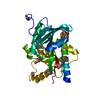
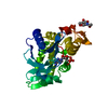
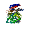
 Structure visualization
 Downloads & links
 Other downloads
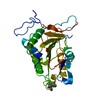


 PDBj
PDBj

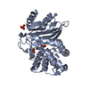
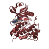
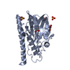
 Assembly
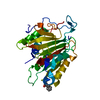


 Mass: 198.260 Da / Num. of mol.: 1 / Source method: obtained synthetically / Formula: C14H14O
 Mass: 18.015 Da / Num. of mol.: 225 / Source method: isolated from a natural source / Formula: H2O
 Sample preparation
 / Beamline: BL41XU / Wavelength: 0.9793 Å
 Processing