- PDB-4exr: Crystal structure of a putative lipoprotein (CD1622) from Clostri... -
+
Open data
ID or keywords:
Loading...
-
Basic information
Entry
Database: PDB / ID: 4exr
Title
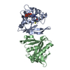
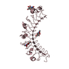
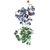
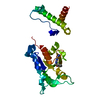
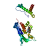
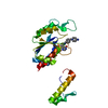
Crystal structure of a putative lipoprotein (CD1622) from Clostridium difficile 630 at 1.85 A resolution
Components
Putative lipoprotein
Keywords
UNKNOWN FUNCTION / YPEB domain dimer / Structural Genomics / Joint Center for Structural Genomics / JCSG / Protein Structure Initiative / PSI-BIOLOGY
Function / homology
Nuclear Transport Factor 2; Chain: A, - #40 / PepSY domain / Peptidase propeptide and YPEB domain / Nuclear Transport Factor 2; Chain: A, / Prokaryotic membrane lipoprotein lipid attachment site profile. / Roll / Alpha Beta / PHOSPHATE ION / Peptidase propeptide and ypeb domain protein
Function and homology information
Biological species
Clostridium difficile (bacteria)
Method
X-RAY DIFFRACTION / SYNCHROTRON / MAD / Resolution: 1.85 Å
Mass: 18.015 Da / Num. of mol.: 104 / Source method: isolated from a natural source / Formula: H2O
Has protein modification
Y
Sequence details
THE CONSTRUCT (RESIDUES 33-205) WAS EXPRESSED WITH A PURIFICATION TAG MGSDKIHHHHHHENLYFQG. THE TAG ...THE CONSTRUCT (RESIDUES 33-205) WAS EXPRESSED WITH A PURIFICATION TAG MGSDKIHHHHHHENLYFQG. THE TAG WAS REMOVED WITH TEV PROTEASE LEAVING ONLY A GLYCINE (0) FOLLOWED BY THE TARGET SEQUENCE.
-
Experimental details
-
Experiment
Experiment
Method: X-RAY DIFFRACTION / Number of used crystals: 1
-
Sample preparation
Crystal
Density Matthews: 3.14 Å3/Da / Density % sol: 60.81 %
Monochromator: single crystal Si(111) bent / Protocol: MAD / Monochromatic (M) / Laue (L): M / Scattering type: x-ray
Radiation wavelength
ID
Wavelength (Å)
Relative weight
1
0.97903
1
2
0.91837
1
3
0.97845
1
Reflection
Resolution: 1.85→28.639 Å / Num. all: 22697 / Num. obs: 22697 / % possible obs: 99.7 % / Redundancy: 5.8 % / Rsym value: 0.093 / Net I/σ(I): 8.7
Reflection shell
Diffraction-ID: 1
Resolution (Å)
Redundancy (%)
Rmerge(I) obs
Mean I/σ(I) obs
Num. measured all
Num. unique all
Rsym value
% possible all
1.85-1.9
5.3
0.817
0.9
8643
1626
0.817
100
1.9-1.95
5.6
0.59
1.2
9024
1624
0.59
99.9
1.95-2.01
6.3
0.459
1.6
9656
1541
0.459
100
2.01-2.07
6.2
0.379
1.9
9437
1531
0.379
100
2.07-2.14
5.9
0.336
2.2
8741
1475
0.336
99.9
2.14-2.21
5.8
0.268
2.8
8322
1436
0.268
99.8
2.21-2.29
5.2
0.228
3.1
7237
1381
0.228
99.6
2.29-2.39
5.8
0.204
3.6
7636
1325
0.204
99.9
2.39-2.49
6.2
0.166
4.4
8022
1290
0.166
100
2.49-2.62
6.1
0.135
5.2
7558
1230
0.135
99.8
2.62-2.76
6
0.12
5.6
7094
1173
0.12
100
2.76-2.93
5.7
0.098
6.6
6280
1108
0.098
99.8
2.93-3.13
5.6
0.086
7.3
5815
1041
0.086
99.5
3.13-3.38
6.3
0.081
7.8
6300
997
0.081
99.9
3.38-3.7
6.1
0.071
8.9
5490
904
0.071
99.5
3.7-4.14
5.7
0.062
10.6
4759
829
0.062
100
4.14-4.78
5.2
0.056
10.7
3834
732
0.056
98.2
4.78-5.85
6
0.059
10.9
3856
639
0.059
100
5.85-8.27
5.2
0.06
11.1
2646
511
0.06
99
8.27-28.639
5.3
0.052
12.2
1618
304
0.052
95.6
-
Phasing
Phasing
Method: MAD
-
Processing
Software
Name
Version
Classification
NB
MolProbity
3beta29
modelbuilding
PDB_EXTRACT
3.1
dataextraction
SOLVE
phasing
SCALA
3.3.20
datascaling
PHENIX
1.7.3
refinement
MOSFLM
datareduction
Refinement
Method to determine structure: MAD / Resolution: 1.85→28.639 Å / Occupancy max: 1 / Occupancy min: 0.5 / SU ML: 0.21 / σ(F): 1.34 / Phase error: 20.78 / Stereochemistry target values: MLHL Details: 1. A MET-INHIBITION PROTOCOL WAS USED FOR SELENOMETHIONINE INCORPORATION DURING PROTEIN EXPRESSION. THE OCCUPANCY OF THE SE ATOMS IN THE MSE RESIDUES WAS REDUCED TO 0.75 FOR THE REDUCED ...Details: 1. A MET-INHIBITION PROTOCOL WAS USED FOR SELENOMETHIONINE INCORPORATION DURING PROTEIN EXPRESSION. THE OCCUPANCY OF THE SE ATOMS IN THE MSE RESIDUES WAS REDUCED TO 0.75 FOR THE REDUCED SCATTERING POWER DUE TO PARTIAL S-MET INCORPORATION. 2. ATOM RECORD CONTAINS SUM OF TLS AND RESIDUAL B FACTORS. ANISOU RECORD CONTAINS SUM OF TLS AND RESIDUAL U FACTORS. 3. THE MAD PHASES WERE USED AS RESTRAINTS DURING REFINEMENT. 4.A SODIUM ION AND PHOSPHATE MOLECULE FROM THE PURIFICATION/CRYSTALLIZATION SOLUTIONS HAVE BEEN MODELED INTO THE STRUCTURE.
Rfactor
Num. reflection
% reflection
Rfree
0.21
1151
5.09 %
Rwork
0.1844
-
-
obs
0.1856
22606
99.35 %
Solvent computation
Shrinkage radii: 0.86 Å / VDW probe radii: 1.1 Å / Solvent model: FLAT BULK SOLVENT MODEL / Bsol: 44.591 Å2 / ksol: 0.364 e/Å3
In the structure databanks used in Yorodumi, some data are registered as the other names, "COVID-19 virus" and "2019-nCoV". Here are the details of the virus and the list of structure data.
Jan 31, 2019. EMDB accession codes are about to change! (news from PDBe EMDB page)
EMDB accession codes are about to change! (news from PDBe EMDB page)
The allocation of 4 digits for EMDB accession codes will soon come to an end. Whilst these codes will remain in use, new EMDB accession codes will include an additional digit and will expand incrementally as the available range of codes is exhausted. The current 4-digit format prefixed with “EMD-” (i.e. EMD-XXXX) will advance to a 5-digit format (i.e. EMD-XXXXX), and so on. It is currently estimated that the 4-digit codes will be depleted around Spring 2019, at which point the 5-digit format will come into force.
The EM Navigator/Yorodumi systems omit the EMD- prefix.
Related info.:Q: What is EMD? / ID/Accession-code notation in Yorodumi/EM Navigator
Yorodumi is a browser for structure data from EMDB, PDB, SASBDB, etc.
This page is also the successor to EM Navigator detail page, and also detail information page/front-end page for Omokage search.
The word "yorodu" (or yorozu) is an old Japanese word meaning "ten thousand". "mi" (miru) is to see.
Related info.:EMDB / PDB / SASBDB / Comparison of 3 databanks / Yorodumi Search / Aug 31, 2016. New EM Navigator & Yorodumi / Yorodumi Papers / Jmol/JSmol / Function and homology information / Changes in new EM Navigator and Yorodumi
 Movie
Movie Controller

 Yorodumi
 Open data
 Basic information
 Components
 Keywords
 Function and homology information
 Clostridium difficile (bacteria)
 X-RAY DIFFRACTION /
  Authors
 Citation
 Structure visualization
 Downloads & links
 Other downloads


 PDBj
PDBj


 Assembly
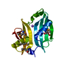


 Mass: 22.990 Da / Num. of mol.: 1 / Source method: obtained synthetically / Formula: Na
 Mass: 94.971 Da / Num. of mol.: 1 / Source method: obtained synthetically / Formula: PO4
 Mass: 18.015 Da / Num. of mol.: 104 / Source method: isolated from a natural source / Formula: H2O
 Sample preparation
 / Beamline: BL11-1 / Wavelength: 0.97903,0.91837,0.97845
 Processing