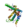
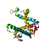
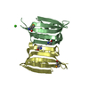
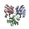
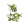
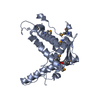
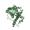
Crystal structure of mouse ryanodine receptor 2 (2699-2904)
Components
Ryanodine receptor 2
Keywords
METAL TRANSPORT / Ryanodine Receptor Calcium Release Channel / Phosphorylation / Muscle / Cardiac
Function / homology
Function and homology information
manganese ion transmembrane transport / establishment of protein localization to endoplasmic reticulum / : / type B pancreatic cell apoptotic process / Purkinje myocyte to ventricular cardiac muscle cell signaling / regulation of atrial cardiac muscle cell action potential / left ventricular cardiac muscle tissue morphogenesis / suramin binding / regulation of AV node cell action potential / sarcoplasmic reticulum calcium ion transport ...manganese ion transmembrane transport / establishment of protein localization to endoplasmic reticulum / : / type B pancreatic cell apoptotic process / Purkinje myocyte to ventricular cardiac muscle cell signaling / regulation of atrial cardiac muscle cell action potential / left ventricular cardiac muscle tissue morphogenesis / suramin binding / regulation of AV node cell action potential / sarcoplasmic reticulum calcium ion transport / regulation of SA node cell action potential / calcium-induced calcium release activity / A band / Stimuli-sensing channels / ventricular cardiac muscle cell action potential / Ion homeostasis / cardiac muscle hypertrophy / embryonic heart tube morphogenesis / regulation of ventricular cardiac muscle cell action potential / calcium ion transport into cytosol / ryanodine-sensitive calcium-release channel activity / release of sequestered calcium ion into cytosol by sarcoplasmic reticulum / response to redox state / regulation of cardiac muscle contraction by calcium ion signaling / cellular response to caffeine / extrinsic component of cytoplasmic side of plasma membrane / response to caffeine / response to muscle activity / calcium ion transmembrane import into cytosol / protein kinase A regulatory subunit binding / protein kinase A catalytic subunit binding / negative regulation of cytosolic calcium ion concentration / positive regulation of the force of heart contraction / smooth endoplasmic reticulum / intracellularly gated calcium channel activity / response to magnesium ion / detection of calcium ion / regulation of cardiac muscle contraction / regulation of cytosolic calcium ion concentration / positive regulation of heart rate / release of sequestered calcium ion into cytosol / response to muscle stretch / cardiac muscle contraction / regulation of cardiac muscle contraction by regulation of the release of sequestered calcium ion / cellular response to epinephrine stimulus / sarcoplasmic reticulum membrane / calcium channel complex / regulation of heart rate / striated muscle contraction / sarcoplasmic reticulum / sarcomere / calcium-mediated signaling / sarcolemma / response to calcium ion / intracellular calcium ion homeostasis / Z disc / calcium channel activity / calcium ion transmembrane transport / calcium ion transport / nuclear envelope / scaffold protein binding / monoatomic ion transmembrane transport / response to hypoxia / calmodulin binding / calcium ion binding / protein kinase binding / enzyme binding / protein-containing complex / membrane / identical protein binding Similarity search - Function
In the structure databanks used in Yorodumi, some data are registered as the other names, "COVID-19 virus" and "2019-nCoV". Here are the details of the virus and the list of structure data.
Jan 31, 2019. EMDB accession codes are about to change! (news from PDBe EMDB page)
EMDB accession codes are about to change! (news from PDBe EMDB page)
The allocation of 4 digits for EMDB accession codes will soon come to an end. Whilst these codes will remain in use, new EMDB accession codes will include an additional digit and will expand incrementally as the available range of codes is exhausted. The current 4-digit format prefixed with “EMD-” (i.e. EMD-XXXX) will advance to a 5-digit format (i.e. EMD-XXXXX), and so on. It is currently estimated that the 4-digit codes will be depleted around Spring 2019, at which point the 5-digit format will come into force.
The EM Navigator/Yorodumi systems omit the EMD- prefix.
Related info.:Q: What is EMD? / ID/Accession-code notation in Yorodumi/EM Navigator
Yorodumi is a browser for structure data from EMDB, PDB, SASBDB, etc.
This page is also the successor to EM Navigator detail page, and also detail information page/front-end page for Omokage search.
The word "yorodu" (or yorozu) is an old Japanese word meaning "ten thousand". "mi" (miru) is to see.
Related info.:EMDB / PDB / SASBDB / Comparison of 3 databanks / Yorodumi Search / Aug 31, 2016. New EM Navigator & Yorodumi / Yorodumi Papers / Jmol/JSmol / Function and homology information / Changes in new EM Navigator and Yorodumi
 Movie
Movie Controller


 Open data
 Basic information
 Components
 Keywords
 Function and homology information
 X-RAY DIFFRACTION /
  Authors
 Citation
 Structure visualization
 Downloads & links
 Other downloads


 PDBj
PDBj


 Assembly
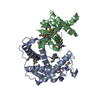


 Mass: 35.453 Da / Num. of mol.: 1 / Mutation: K2879A / Source method: obtained synthetically / Formula: Cl
 Mass: 18.015 Da / Num. of mol.: 329 / Source method: isolated from a natural source / Formula: H2O
 Sample preparation
 / Beamline: 08ID-1 / Wavelength: 0.9795 Å
 Processing