- PDB-4esn: Crystal structure of a DUF1312 family protein (RUMGNA_02503) from... -
+
Open data
ID or keywords:
Loading...
-
Basic information
Entry
Database: PDB / ID: 4esn
Title
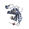
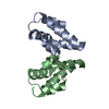
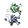
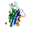
Crystal structure of a DUF1312 family protein (RUMGNA_02503) from Ruminococcus gnavus ATCC 29149 at 2.20 A resolution
Components
hypothetical protein
Keywords
Structural Genomics / Unknown Function / PROTEIN OF PF07009 FAMILY / DUF1312 / JOINT CENTER FOR STRUCTURAL GENOMICS / JCSG / PROTEIN STRUCTURE INITIATIVE / PSI-BIOLOGY
Function / homology
NusG, domain 2 / N-utilization substance G protein NusG, insert domain / NusG, domain 2 superfamily / NusG domain II / mini-chromosome maintenance (MCM) complex, domain 2 / Sandwich / Mainly Beta / metal ion binding / NusG domain-containing protein
Function and homology information
Biological species
Ruminococcus gnavus (bacteria)
Method
X-RAY DIFFRACTION / SYNCHROTRON / SAD / Resolution: 2.2 Å
Mass: 18.015 Da / Num. of mol.: 53 / Source method: isolated from a natural source / Formula: H2O
Has protein modification
Y
Sequence details
THE CONSTRUCT WAS EXPRESSED WITH AN N-TERMINAL PURIFICATION TAG MGSDKIHHHHHHENLYFQG. THE TAG WAS ...THE CONSTRUCT WAS EXPRESSED WITH AN N-TERMINAL PURIFICATION TAG MGSDKIHHHHHHENLYFQG. THE TAG WAS REMOVED WITH TEV PROTEASE LEAVING ONLY A GLYCINE (0) FOLLOWED BY RESIDUES 33-117 OF THE TARGET SEQUENCE. ANALYSIS OF THE PURIFIED PROTEIN BY MASS SPECTROMETRY AND GEL ELECTROPHORESIS SHOWS THAT WHILE THE MAJORITY OF THE PROTEIN WAS CLEAVED, THERE WAS SOME UNCLEAVED PROTEIN PRESENT. SINCE ELECTRON DENSITY WAS OBSERVED FOR RESIDUES -2 AND -1 IN BOTH CHAINS, THE TAG SEQUENCE IS INCLUDED IN THE SEQRES RECORDS. THE CRYSTAL MAY CONTAIN A MIXTURE OF TAG-ON AND TAG-OFF PROTEIN.
-
Experimental details
-
Experiment
Experiment
Method: X-RAY DIFFRACTION / Number of used crystals: 1
-
Sample preparation
Crystal
Density Matthews: 1.82 Å3/Da / Density % sol: 32.33 %
Crystal grow
Temperature: 293 K / Method: vapor diffusion, sitting drop Details: 0.2M ammonium nitrate 20.0% polyethylene glycol 3350, NANODROP, VAPOR DIFFUSION, SITTING DROP, temperature 293K
Type: MARMOSAIC 325 mm CCD / Detector: CCD / Date: Jan 13, 2012 / Details: double crystal monochromator
Radiation
Monochromator: double crystal / Protocol: SINGLE WAVELENGTH / Monochromatic (M) / Laue (L): M / Scattering type: x-ray
Radiation wavelength
Wavelength: 0.97915 Å / Relative weight: 1
Reflection
Resolution: 2.2→28.58 Å / Num. all: 9044 / Num. obs: 9044 / % possible obs: 99.5 % / Redundancy: 7.1 % / Rsym value: 0.081 / Net I/σ(I): 11.5
Reflection shell
Diffraction-ID: 1
Resolution (Å)
Redundancy (%)
Rmerge(I) obs
Mean I/σ(I) obs
Num. measured all
Num. unique all
Rsym value
% possible all
2.2-2.26
7.3
0.695
1.1
4772
656
0.695
99
2.26-2.32
7.3
0.566
1.4
4532
625
0.566
99.7
2.32-2.39
7.3
0.514
1.5
4525
621
0.514
99.4
2.39-2.46
7.3
0.375
2
4313
592
0.375
99.2
2.46-2.54
7.2
0.323
2.4
4252
591
0.323
99.4
2.54-2.63
7.2
0.25
3.1
4056
560
0.25
99.6
2.63-2.73
7.2
0.183
4.1
3989
551
0.183
99.4
2.73-2.84
7.2
0.152
4.8
3855
537
0.152
99.8
2.84-2.97
7.2
0.133
5.4
3633
505
0.133
99.7
2.97-3.11
7.1
0.117
5.8
3503
493
0.117
99.7
3.11-3.28
7.2
0.091
7
3419
475
0.091
99.8
3.28-3.48
7.1
0.086
7.3
3130
441
0.086
99.8
3.48-3.72
7
0.076
8.4
2973
423
0.076
99.8
3.72-4.02
7
0.07
8.8
2792
398
0.07
99.7
4.02-4.4
7
0.059
10.1
2543
364
0.059
99.9
4.4-4.92
6.9
0.06
10.6
2286
331
0.06
99.8
4.92-5.68
6.8
0.066
10
2040
302
0.066
99.9
5.68-6.96
6.6
0.079
8.8
1673
253
0.079
100
6.96-9.84
6.3
0.056
10.6
1336
212
0.056
99.3
9.84-28.58
5.5
0.061
10.2
626
114
0.061
90.3
-
Phasing
Phasing
Method: SAD
-
Processing
Software
Name
Version
Classification
NB
MolProbity
3beta29
modelbuilding
PDB_EXTRACT
3.1
dataextraction
SHELX
phasing
SHARP
phasing
SCALA
3.3.20
datascaling
BUSTER-TNT
2.10.0
refinement
MOSFLM
datareduction
SHELXD
phasing
BUSTER
2.10.0
refinement
Refinement
Method to determine structure: SAD / Resolution: 2.2→28.58 Å / Cor.coef. Fo:Fc: 0.9552 / Cor.coef. Fo:Fc free: 0.9477 / Occupancy max: 1 / Occupancy min: 0.5 / Cross valid method: THROUGHOUT / σ(F): 0 Details: 1. ATOM RECORD CONTAINS SUM OF TLS AND RESIDUAL B FACTORS. ANISOU RECORD CONTAINS SUM OF TLS AND RESIDUAL U FACTORS. 2. A MET-INHIBITION PROTOCOL WAS USED FOR SELENOMETHIONINE INCORPORATION ...Details: 1. ATOM RECORD CONTAINS SUM OF TLS AND RESIDUAL B FACTORS. ANISOU RECORD CONTAINS SUM OF TLS AND RESIDUAL U FACTORS. 2. A MET-INHIBITION PROTOCOL WAS USED FOR SELENOMETHIONINE INCORPORATION DURING PROTEIN EXPRESSION. THE OCCUPANCY OF THE SE ATOMS IN THE MSE RESIDUES WAS REDUCED TO 0.75 TO ACCOUNT FOR THE REDUCED SCATTERING POWER DUE TO PARTIAL S-MET INCORPORATION. 3. POLYETHYLENE GLYCOL (PE4) FROM THE CRYOPROTECTANT AND CL IONS FROM THE CRYSTALLIZATION CONDITION HAVE BEEN MODELED IN THE SOLVENT STRUCTURE. 4. ZN ION WAS MODELED IN THE PUTATIVE ACTIVE CENTER OF EACH PROTOME BASED ON ANOMALOUS DIFFERENCE MAPS AND EXCITATION SCANS. 5. 11 C-TERMINAL RESIDUES OF A AND B MOLECULES WERE DISORDERED. 6. NCS RESTRAINTS WERE APPLIED USING BUSTER'S LSSR RESTRAINT REPRESENTATION (-AUTONCS).
In the structure databanks used in Yorodumi, some data are registered as the other names, "COVID-19 virus" and "2019-nCoV". Here are the details of the virus and the list of structure data.
Jan 31, 2019. EMDB accession codes are about to change! (news from PDBe EMDB page)
EMDB accession codes are about to change! (news from PDBe EMDB page)
The allocation of 4 digits for EMDB accession codes will soon come to an end. Whilst these codes will remain in use, new EMDB accession codes will include an additional digit and will expand incrementally as the available range of codes is exhausted. The current 4-digit format prefixed with “EMD-” (i.e. EMD-XXXX) will advance to a 5-digit format (i.e. EMD-XXXXX), and so on. It is currently estimated that the 4-digit codes will be depleted around Spring 2019, at which point the 5-digit format will come into force.
The EM Navigator/Yorodumi systems omit the EMD- prefix.
Related info.:Q: What is EMD? / ID/Accession-code notation in Yorodumi/EM Navigator
Yorodumi is a browser for structure data from EMDB, PDB, SASBDB, etc.
This page is also the successor to EM Navigator detail page, and also detail information page/front-end page for Omokage search.
The word "yorodu" (or yorozu) is an old Japanese word meaning "ten thousand". "mi" (miru) is to see.
Related info.:EMDB / PDB / SASBDB / Comparison of 3 databanks / Yorodumi Search / Aug 31, 2016. New EM Navigator & Yorodumi / Yorodumi Papers / Jmol/JSmol / Function and homology information / Changes in new EM Navigator and Yorodumi
 Movie
Movie Controller

 Yorodumi
 Open data
 Basic information
 Components
 Keywords
 Function and homology information
 Ruminococcus gnavus (bacteria)
 X-RAY DIFFRACTION /
  Authors
 Citation
 Structure visualization
 Downloads & links
 Other downloads
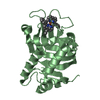


 PDBj
PDBj Assembly
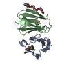

 Mass: 65.409 Da / Num. of mol.: 2 / Source method: obtained synthetically / Formula: Zn
 Mass: 354.436 Da / Num. of mol.: 1 / Source method: obtained synthetically / Formula: C16H34O8 / Comment: precipitant*YM
 Mass: 18.015 Da / Num. of mol.: 53 / Source method: isolated from a natural source / Formula: H2O
 Sample preparation
 / Beamline: BL9-2 / Wavelength: 0.97915
  Processing