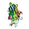
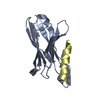
[English] 日本語
Yorodumi
- PDB-4l1l: Rat PKC C2 domain bound to CD -

+
Open data


ID or keywords:

Loading...

-
Basic information

Entry
Database: PDB / ID: 4l1l
TitleRat PKC C2 domain bound to CD
ComponentsProtein kinase C alpha type
KeywordsTRANSFERASE / PROTEIN KINASE PKC
Function / homology
Function and homology information


Acetylcholine regulates insulin secretion / EGFR Transactivation by Gastrin / HuR (ELAVL1) binds and stabilizes mRNA / Depolymerization of the Nuclear Lamina / Signaling by SCF-KIT / Regulation of KIT signaling / Calmodulin induced events / Disinhibition of SNARE formation / SHC1 events in ERBB2 signaling / Response to elevated platelet cytosolic Ca2+ ...Acetylcholine regulates insulin secretion / EGFR Transactivation by Gastrin / HuR (ELAVL1) binds and stabilizes mRNA / Depolymerization of the Nuclear Lamina / Signaling by SCF-KIT / Regulation of KIT signaling / Calmodulin induced events / Disinhibition of SNARE formation / SHC1 events in ERBB2 signaling / Response to elevated platelet cytosolic Ca2+ / Syndecan interactions / positive regulation of angiotensin-activated signaling pathway / cellular response to carbohydrate stimulus / response to phorbol 13-acetate 12-myristate / positive regulation of dense core granule biogenesis / VEGFR2 mediated cell proliferation / cone photoreceptor outer segment / calcium,diacylglycerol-dependent serine/threonine kinase activity / protein kinase C signaling / ooplasm / RET signaling / central nervous system neuron axonogenesis / desmosome assembly / WNT5A-dependent internalization of FZD4 / RHO GTPases Activate NADPH Oxidases / protein kinase C / diacylglycerol-dependent serine/threonine kinase activity / negative regulation of glial cell apoptotic process / regulation of platelet aggregation / positive regulation of macrophage differentiation / negative regulation of D-glucose import / regulation of muscle contraction / regulation of the force of heart contraction / induction of positive chemotaxis / alphav-beta3 integrin-PKCalpha complex / positive regulation of lipopolysaccharide-mediated signaling pathway / positive regulation of synapse assembly / response to corticosterone / muscle cell cellular homeostasis / positive regulation of cardiac muscle hypertrophy / regulation of synaptic vesicle exocytosis / Trafficking of GluR2-containing AMPA receptors / positive regulation of exocytosis / positive regulation of bone resorption / intercalated disc / peptidyl-threonine phosphorylation / positive regulation of blood vessel endothelial cell migration / response to mechanical stimulus / chondrocyte differentiation / presynaptic cytosol / negative regulation of MAPK cascade / positive regulation of endothelial cell proliferation / neutrophil chemotaxis / presynaptic modulation of chemical synaptic transmission / positive regulation of endothelial cell migration / positive regulation of cell adhesion / positive regulation of mitotic cell cycle / response to interleukin-1 / intrinsic apoptotic signaling pathway / negative regulation of insulin receptor signaling pathway / calyx of Held / response to reactive oxygen species / histone H3T6 kinase activity / positive regulation of smooth muscle cell proliferation / stem cell differentiation / establishment of protein localization / mitochondrial membrane / response to peptide hormone / response to toxic substance / intracellular calcium ion homeostasis / positive regulation of inflammatory response / positive regulation of angiogenesis / integrin binding / apical part of cell / response to estradiol / angiogenesis / response to ethanol / learning or memory / cell population proliferation / positive regulation of ERK1 and ERK2 cascade / protein kinase activity / cell adhesion / negative regulation of translation / intracellular signal transduction / ciliary basal body / positive regulation of cell migration / axon / signaling receptor binding / negative regulation of cell population proliferation / protein serine kinase activity / neuronal cell body / protein serine/threonine kinase activity / lipid binding / dendrite / perinuclear region of cytoplasm / enzyme binding / endoplasmic reticulum / protein-containing complex / mitochondrion / zinc ion binding
Similarity search - Function
Classical Protein Kinase C alpha, catalytic domain / Protein kinase C, alpha/beta/gamma types / Protein kinase, C-terminal / Protein kinase C terminal domain / Diacylglycerol/phorbol-ester binding / Phorbol esters/diacylglycerol binding domain (C1 domain) / C2 domain / Zinc finger phorbol-ester/DAG-type signature. / Protein kinase C conserved region 2 (CalB) / C2 domain ...Classical Protein Kinase C alpha, catalytic domain / Protein kinase C, alpha/beta/gamma types / Protein kinase, C-terminal / Protein kinase C terminal domain / Diacylglycerol/phorbol-ester binding / Phorbol esters/diacylglycerol binding domain (C1 domain) / C2 domain / Zinc finger phorbol-ester/DAG-type signature. / Protein kinase C conserved region 2 (CalB) / C2 domain / Zinc finger phorbol-ester/DAG-type profile. / Protein kinase C conserved region 1 (C1) domains (Cysteine-rich domains) / Protein kinase C-like, phorbol ester/diacylglycerol-binding domain / C2 domain / C2 domain profile. / C1-like domain superfamily / Extension to Ser/Thr-type protein kinases / AGC-kinase, C-terminal / AGC-kinase C-terminal domain profile. / C2 domain superfamily / Serine/threonine-protein kinase, active site / Serine/Threonine protein kinases active-site signature. / Protein kinase domain / Serine/Threonine protein kinases, catalytic domain / Protein kinase, ATP binding site / Protein kinases ATP-binding region signature. / Protein kinase domain profile. / Protein kinase domain / Protein kinase-like domain superfamily / Immunoglobulin-like / Sandwich / Mainly Beta
Similarity search - Domain/homology
: / Protein kinase C alpha type
Similarity search - Component
Biological speciesRattus norvegicus (Norway rat)
MethodX-RAY DIFFRACTION / SAD / Resolution: 1.6 Å
AuthorsMorales, K.M. / Yang, Y. / Long, Z. / Li, P. / Taylor, A.B. / Hart, P.J. / Igumenova, T.I.
CitationJournal: J.Am.Chem.Soc. / Year: 2013
Title: Cd(2+) as a ca(2+) surrogate in protein-membrane interactions: isostructural but not isofunctional.
Authors: Morales, K.A. / Yang, Y. / Long, Z. / Li, P. / Taylor, A.B. / Hart, P.J. / Igumenova, T.I.
History
DepositionJun 3, 2013Deposition site: RCSB / Processing site: RCSB
Revision 1.0Aug 28, 2013Provider: repository / Type: Initial release
Revision 1.1Oct 23, 2013Group: Database references
Revision 1.2Feb 28, 2024Group: Data collection / Database references / Derived calculations
Category: chem_comp_atom / chem_comp_bond ...chem_comp_atom / chem_comp_bond / database_2 / pdbx_struct_conn_angle / struct_conn / struct_site
Item: _database_2.pdbx_DOI / _database_2.pdbx_database_accession ..._database_2.pdbx_DOI / _database_2.pdbx_database_accession / _pdbx_struct_conn_angle.ptnr1_auth_comp_id / _pdbx_struct_conn_angle.ptnr1_auth_seq_id / _pdbx_struct_conn_angle.ptnr1_label_alt_id / _pdbx_struct_conn_angle.ptnr1_label_asym_id / _pdbx_struct_conn_angle.ptnr1_label_atom_id / _pdbx_struct_conn_angle.ptnr1_label_comp_id / _pdbx_struct_conn_angle.ptnr1_label_seq_id / _pdbx_struct_conn_angle.ptnr2_auth_seq_id / _pdbx_struct_conn_angle.ptnr2_label_asym_id / _pdbx_struct_conn_angle.ptnr3_auth_comp_id / _pdbx_struct_conn_angle.ptnr3_auth_seq_id / _pdbx_struct_conn_angle.ptnr3_label_alt_id / _pdbx_struct_conn_angle.ptnr3_label_asym_id / _pdbx_struct_conn_angle.ptnr3_label_atom_id / _pdbx_struct_conn_angle.ptnr3_label_comp_id / _pdbx_struct_conn_angle.ptnr3_label_seq_id / _pdbx_struct_conn_angle.value / _struct_conn.pdbx_dist_value / _struct_conn.pdbx_ptnr1_label_alt_id / _struct_conn.ptnr1_auth_comp_id / _struct_conn.ptnr1_auth_seq_id / _struct_conn.ptnr1_label_asym_id / _struct_conn.ptnr1_label_atom_id / _struct_conn.ptnr1_label_comp_id / _struct_conn.ptnr1_label_seq_id / _struct_conn.ptnr2_auth_comp_id / _struct_conn.ptnr2_auth_seq_id / _struct_conn.ptnr2_label_asym_id / _struct_conn.ptnr2_label_atom_id / _struct_conn.ptnr2_label_comp_id / _struct_site.pdbx_auth_asym_id / _struct_site.pdbx_auth_comp_id / _struct_site.pdbx_auth_seq_id

-
Structure visualization

Structure viewerMolecule:
MolmilJmol/JSmol

Downloads & links

-
Assembly

Deposited unit
A: Protein kinase C alpha type
hetero molecules


Theoretical massNumber of molelcules
Total (without water)17,39512
Polymers16,2401
Non-polymers1,15511
Water2,540141
1


  • Idetical with deposited unit
  • defined by author&software
TypeNameSymmetry operationNumber
identity operation1_555x,y,z1
MethodPISA
Unit cell
Length a, b, c (Å)58.224, 58.224, 88.539
Angle α, β, γ (deg.)90.00, 90.00, 120.00
Int Tables number154
Space group name H-MP3221
Components on special symmetry positions
IDModelComponents
11A-429-

HOH

21A-478-

HOH

-
Components

#1: Protein Protein kinase C alpha type / PKC-A / PKC-alpha


Mass: 16240.451 Da / Num. of mol.: 1 / Fragment: UNP RESIDUES 155-293
Source method: isolated from a genetically manipulated source
Source: (gene. exp.) Rattus norvegicus (Norway rat) / Gene: Pkca, Prkca / Plasmid: pET-SUMO / Production host: Escherichia coli (E. coli) / References: UniProt: P05696, protein kinase C
#2: Chemical
ChemComp-SO4 / SULFATE ION


Mass: 96.063 Da / Num. of mol.: 5 / Source method: obtained synthetically / Formula: SO4
#3: Chemical
ChemComp-CD / CADMIUM ION


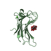
Mass: 112.411 Da / Num. of mol.: 6 / Source method: obtained synthetically / Formula: Cd
#4: Water ChemComp-HOH / water


Mass: 18.015 Da / Num. of mol.: 141 / Source method: isolated from a natural source / Formula: H2O

-
Experimental details

-
Experiment

ExperimentMethod: X-RAY DIFFRACTION / Number of used crystals: 1

-
Sample preparation

CrystalDensity Matthews: 2.67 Å3/Da / Density % sol: 53.89 %
Crystal growTemperature: 298 K / Method: vapor diffusion, hanging drop / pH: 5.25
Details: 0.25 M Lithium Sulfate, 20% PEG 3350, pH 5.25, VAPOR DIFFUSION, HANGING DROP, temperature 298K

-
Data collection

DiffractionMean temperature: 100 K
Diffraction sourceSource: ROTATING ANODE / Type: RIGAKU MICROMAX-007 HF / Wavelength: 1.54178 Å
DetectorType: RIGAKU RAXIS IV++ / Detector: IMAGE PLATE / Date: Mar 25, 2013
RadiationProtocol: SINGLE WAVELENGTH / Monochromatic (M) / Laue (L): M / Scattering type: x-ray
Radiation wavelengthWavelength: 1.54178 Å / Relative weight: 1
ReflectionResolution: 1.6→44.27 Å / Num. obs: 43697 / % possible obs: 98.6 % / Redundancy: 20 % / Biso Wilson estimate: 20.02 Å2 / Rsym value: 0.098 / Net I/σ(I): 22.4
Reflection shellResolution: 1.6→1.69 Å / Redundancy: 17.8 % / Mean I/σ(I) obs: 7.2 / Num. unique all: 3157 / Rsym value: 0.397 / % possible all: 94.3

-
Processing

Software
NameVersionClassification
PHENIX(phenix.refine: 1.8.2_1309)refinement
XDSdata reduction
XDSdata scaling
RefinementMethod to determine structure: SAD / Resolution: 1.6→43.816 Å / Isotropic thermal model: anisotropic / Cross valid method: THROUGHOUT / σ(F): 1.7 / Phase error: 17.9 / Stereochemistry target values: ML
RfactorNum. reflection% reflectionSelection details
Rfree0.1953 2000 8.67 %random
Rwork0.1568 ---
obs0.16 43697 98.5 %-
Solvent computationShrinkage radii: 0.8 Å / VDW probe radii: 1 Å / Solvent model: FLAT BULK SOLVENT MODEL
Displacement parametersBiso mean: 27.09 Å2
Refinement stepCycle: LAST / Resolution: 1.6→43.816 Å
ProteinNucleic acidLigandSolventTotal
Num. atoms1133 0 31 141 1305
Refine LS restraints
Refine-IDTypeDev idealNumber
X-RAY DIFFRACTIONf_bond_d0.011224
X-RAY DIFFRACTIONf_angle_d1.2251664
X-RAY DIFFRACTIONf_dihedral_angle_d13.911480
X-RAY DIFFRACTIONf_chiral_restr0.087171
X-RAY DIFFRACTIONf_plane_restr0.007214
LS refinement shell
Resolution (Å)Rfactor RfreeNum. reflection RfreeRfactor RworkNum. reflection RworkRefine-ID% reflection obs (%)
1.5995-1.61980.28661290.2041299X-RAY DIFFRACTION85
1.6198-1.64110.22971340.15511380X-RAY DIFFRACTION96
1.6411-1.66360.20661400.13971533X-RAY DIFFRACTION98
1.6636-1.68730.2241320.13731407X-RAY DIFFRACTION97
1.6873-1.71250.19921460.14061552X-RAY DIFFRACTION98
1.7125-1.73930.16761320.12941422X-RAY DIFFRACTION98
1.7393-1.76780.17221520.12481506X-RAY DIFFRACTION98
1.7678-1.79830.2031340.11991424X-RAY DIFFRACTION99
1.7983-1.8310.18111440.13211508X-RAY DIFFRACTION98
1.831-1.86620.21181420.13581474X-RAY DIFFRACTION99
1.8662-1.90430.20311420.13181451X-RAY DIFFRACTION99
1.9043-1.94570.16691460.12991527X-RAY DIFFRACTION99
1.9457-1.9910.19711360.12311433X-RAY DIFFRACTION99
1.991-2.04070.15651460.13191509X-RAY DIFFRACTION99
2.0407-2.09590.17171400.13091516X-RAY DIFFRACTION99
2.0959-2.15760.19831440.14471459X-RAY DIFFRACTION99
2.1576-2.22720.21781440.14091494X-RAY DIFFRACTION100
2.2272-2.30680.17861440.14961514X-RAY DIFFRACTION100
2.3068-2.39920.22351360.15691518X-RAY DIFFRACTION100
2.3992-2.50840.19491440.15361451X-RAY DIFFRACTION100
2.5084-2.64060.20461380.17221515X-RAY DIFFRACTION100
2.6406-2.8060.221420.17551488X-RAY DIFFRACTION100
2.806-3.02260.20191520.1751502X-RAY DIFFRACTION100
3.0226-3.32670.17781380.16291500X-RAY DIFFRACTION100
3.3267-3.80790.19021440.16281501X-RAY DIFFRACTION100
3.8079-4.79650.1921350.15641524X-RAY DIFFRACTION100
4.7965-43.83250.19731310.18661503X-RAY DIFFRACTION100

+
About Yorodumi

-
News

-
Feb 9, 2022. New format data for meta-information of EMDB entries

New format data for meta-information of EMDB entries

  • Version 3 of the EMDB header file is now the official format.
  • The previous official version 1.9 will be removed from the archive.

Related info.:EMDB header

External links:wwPDB to switch to version 3 of the EMDB data model

-
Aug 12, 2020. Covid-19 info

Covid-19 info

URL: https://pdbj.org/emnavi/covid19.php

New page: Covid-19 featured information page in EM Navigator.

Related info.:Covid-19 info / Mar 5, 2020. Novel coronavirus structure data

+
Mar 5, 2020. Novel coronavirus structure data

Novel coronavirus structure data

Related info.:Yorodumi Speices / Aug 12, 2020. Covid-19 info

External links:COVID-19 featured content - PDBj / Molecule of the Month (242):Coronavirus Proteases

+
Jan 31, 2019. EMDB accession codes are about to change! (news from PDBe EMDB page)

EMDB accession codes are about to change! (news from PDBe EMDB page)

  • The allocation of 4 digits for EMDB accession codes will soon come to an end. Whilst these codes will remain in use, new EMDB accession codes will include an additional digit and will expand incrementally as the available range of codes is exhausted. The current 4-digit format prefixed with “EMD-” (i.e. EMD-XXXX) will advance to a 5-digit format (i.e. EMD-XXXXX), and so on. It is currently estimated that the 4-digit codes will be depleted around Spring 2019, at which point the 5-digit format will come into force.
  • The EM Navigator/Yorodumi systems omit the EMD- prefix.

Related info.:Q: What is EMD? / ID/Accession-code notation in Yorodumi/EM Navigator

External links:EMDB Accession Codes are Changing Soon! / Contact to PDBj

+
Jul 12, 2017. Major update of PDB

Major update of PDB

  • wwPDB released updated PDB data conforming to the new PDBx/mmCIF dictionary.
  • This is a major update changing the version number from 4 to 5, and with Remediation, in which all the entries are updated.
  • In this update, many items about electron microscopy experimental information are reorganized (e.g. em_software).
  • Now, EM Navigator and Yorodumi are based on the updated data.

External links:wwPDB Remediation / Enriched Model Files Conforming to OneDep Data Standards Now Available in the PDB FTP Archive

-
Yorodumi

Thousand views of thousand structures

  • Yorodumi is a browser for structure data from EMDB, PDB, SASBDB, etc.
  • This page is also the successor to EM Navigator detail page, and also detail information page/front-end page for Omokage search.
  • The word "yorodu" (or yorozu) is an old Japanese word meaning "ten thousand". "mi" (miru) is to see.

Related info.:EMDB / PDB / SASBDB / Comparison of 3 databanks / Yorodumi Search / Aug 31, 2016. New EM Navigator & Yorodumi / Yorodumi Papers / Jmol/JSmol / Function and homology information / Changes in new EM Navigator and Yorodumi

Read more