 Movie
Movie Controller

[English] 日本語
 Yorodumi
- PDB-4esa: X-ray structure of carbonmonoxy hemoglobin of Eleginops maclovinus -
+ Open data

- Basic information
| Entry | Database: PDB / ID: 4esa | ||||||
|---|---|---|---|---|---|---|---|
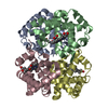
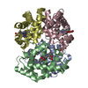
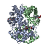
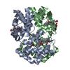
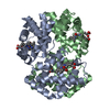
| Title | X-ray structure of carbonmonoxy hemoglobin of Eleginops maclovinus | ||||||
 Components Components |
| ||||||
 Keywords Keywords | OXYGEN TRANSPORT / haemoglobin / ligand-binding properties / Root effect / quaternary structure / structure/function relationship / oxygen transporter / O2 / blood | ||||||
| Function / homology |  Function and homology information Function and homology informationhaptoglobin binding / organic acid binding / haptoglobin-hemoglobin complex / hemoglobin complex / hydrogen peroxide catabolic process / oxygen carrier activity / peroxidase activity / oxygen binding / blood microparticle / iron ion binding ...haptoglobin binding / organic acid binding / haptoglobin-hemoglobin complex / hemoglobin complex / hydrogen peroxide catabolic process / oxygen carrier activity / peroxidase activity / oxygen binding / blood microparticle / iron ion binding / heme binding / metal ion binding Similarity search - Function | ||||||
| Biological species |  Eleginops maclovinus (Patagonian blennie) Eleginops maclovinus (Patagonian blennie) | ||||||
| Method |  X-RAY DIFFRACTION / SYNCHROTRON / MOLECULAR REPLACEMENT / Resolution: 1.45 Å X-RAY DIFFRACTION / SYNCHROTRON / MOLECULAR REPLACEMENT / Resolution: 1.45 Å | ||||||
 Authors Authors | Merlino, A. / Vitagliano, L. / Mazzarella, L. / Vergara, A. | ||||||
 Citation Citation | Journal: Mol Biosyst / Year: 2012 Title: ATP regulation of the ligand-binding properties in temperate and cold-adapted haemoglobins. X-ray structure and ligand-binding kinetics in the sub-Antarctic fish Eleginops maclovinus. Authors: Coppola, D. / Abbruzzetti, S. / Nicoletti, F. / Merlino, A. / Gambacurta, A. / Giordano, D. / Howes, B.D. / De Sanctis, G. / Vitagliano, L. / Bruno, S. / di Prisco, G. / Mazzarella, L. / ...Authors: Coppola, D. / Abbruzzetti, S. / Nicoletti, F. / Merlino, A. / Gambacurta, A. / Giordano, D. / Howes, B.D. / De Sanctis, G. / Vitagliano, L. / Bruno, S. / di Prisco, G. / Mazzarella, L. / Smulevich, G. / Coletta, M. / Viappiani, C. / Vergara, A. / Verde, C. #1: Journal: Biopolymers / Year: 2009Title: Combined crystallographic and spectroscopic analysis of Trematomus bernacchii hemoglobin highlights analogies and differences in the peculiar oxidation pathway of Antarctic fish hemoglobins. Authors: Merlino, A. / Vitagliano, L. / Howes, B.D. / Verde, C. / di Prisco, G. / Smulevich, G. / Sica, F. / Vergara, A. #2: Journal: J.Am.Chem.Soc. / Year: 2008Title: Spectroscopic and crystallographic characterization of a tetrameric hemoglobin oxidation reveals structural features of the functional intermediate relaxed/tense state. Authors: Vitagliano, L. / Vergara, A. / Bonomi, G. / Merlino, A. / Verde, C. / di Prisco, G. / Howes, B.D. / Smulevich, G. / Mazzarella, L. | ||||||
| History |
|
- Structure visualization
| Structure viewer | Molecule: MolmilJmol/JSmol |
|---|
- Downloads & links
-Download
| PDBx/mmCIF format | 4esa.cif.gz | 137.3 KB | Display | PDBx/mmCIF format |
|---|---|---|---|---|
| PDB format | pdb4esa.ent.gz | 107.3 KB | Display | PDB format |
| PDBx/mmJSON format | 4esa.json.gz | Tree view | PDBx/mmJSON format | |
| Others |  Other downloads Other downloads |
-Validation report
| Arichive directory | https://data.pdbj.org/pub/pdb/validation_reports/es/4esaftp://data.pdbj.org/pub/pdb/validation_reports/es/4esa | HTTPS FTP |
|---|
-Related structure data
| Related structure data |  3gkvS S: Starting model for refinement |
|---|---|
| Similar structure data |


-Links
 PDBj
PDBj


- Assembly
| Deposited unit | 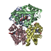
| ||||||||
|---|---|---|---|---|---|---|---|---|---|
| 1 |
| ||||||||
| Unit cell |
|
-Components
-Protein , 2 types, 4 molecules ACBD
| #1: Protein | Mass: 15686.250 Da / Num. of mol.: 2 / Source method: isolated from a natural source / Source: (natural) Eleginops maclovinus (Patagonian blennie) / References: UniProt: K7N5M5*PLUS#2: Protein | Mass: 16153.450 Da / Num. of mol.: 2 / Source method: isolated from a natural source / Source: (natural) Eleginops maclovinus (Patagonian blennie) / References: UniProt: K7N5M6*PLUS |
|---|
-Non-polymers , 4 types, 417 molecules 


| #3: Chemical | ChemComp-CMO /  Mass: 28.010 Da / Num. of mol.: 4 / Source method: obtained synthetically / Formula: CO Mass: 28.010 Da / Num. of mol.: 4 / Source method: obtained synthetically / Formula: CO#4: Chemical | ChemComp-HEM /  Mass: 616.487 Da / Num. of mol.: 4 / Source method: obtained synthetically / Formula: C34H32FeN4O4 Mass: 616.487 Da / Num. of mol.: 4 / Source method: obtained synthetically / Formula: C34H32FeN4O4#5: Chemical | ChemComp-GOL / |  Mass: 92.094 Da / Num. of mol.: 1 / Source method: obtained synthetically / Formula: C3H8O3 Mass: 92.094 Da / Num. of mol.: 1 / Source method: obtained synthetically / Formula: C3H8O3#6: Water | ChemComp-HOH / | Mass: 18.015 Da / Num. of mol.: 408 / Source method: isolated from a natural source / Formula: H2O |
|---|
-Details
| Has protein modification | Y |
|---|
-Experimental details
-Experiment
| Experiment | Method: X-RAY DIFFRACTION / Number of used crystals: 1 |
|---|
- Sample preparation
| Crystal | Density Matthews: 2.48 Å3/Da / Density % sol: 50.36 % |
|---|---|
| Crystal grow | Temperature: 298 K / Method: microdialysis / pH: 8 Details: 10 ml protein solution at 20 mg/ml 1 in 100 mM Tris HCl buffer and 2 mM sodium dithionite was dialyzed against a 25 ml reservoir containing 2 M ammonium sulfate, 100 mM Tris HCl, 2 mM sodium ...Details: 10 ml protein solution at 20 mg/ml 1 in 100 mM Tris HCl buffer and 2 mM sodium dithionite was dialyzed against a 25 ml reservoir containing 2 M ammonium sulfate, 100 mM Tris HCl, 2 mM sodium dithionite, pH 8.0, MICRODIALYSIS, temperature 298K |
-Data collection
| Diffraction | Mean temperature: 100 K |
|---|---|
| Diffraction source | Source: SYNCHROTRON / Site: ELETTRA  / Beamline: 5.2R / Wavelength: 1 Å / Beamline: 5.2R / Wavelength: 1 Å |
| Detector | Type: MAR CCD 165 mm / Detector: CCD / Date: Jul 4, 2009 / Details: mirrors |
| Radiation | Monochromator: graphite / Protocol: SINGLE WAVELENGTH / Monochromatic (M) / Laue (L): M / Scattering type: x-ray |
| Radiation wavelength | Wavelength: 1 Å / Relative weight: 1 |
| Reflection | Resolution: 1.45→50 Å / Num. all: 109738 / Num. obs: 109671 / % possible obs: 97.3 % / Observed criterion σ(F): 0 / Observed criterion σ(I): 0 / Redundancy: 3.5 % / Rmerge(I) obs: 0.075 / Net I/σ(I): 37.5 |
| Reflection shell | Resolution: 1.45→1.5 Å / Redundancy: 2.5 % / Rmerge(I) obs: 0.382 / Mean I/σ(I) obs: 3.3 / % possible all: 90.8 |
- Processing
| Software |
| ||||||||||||||||||||
|---|---|---|---|---|---|---|---|---|---|---|---|---|---|---|---|---|---|---|---|---|---|
| Refinement | Method to determine structure: MOLECULAR REPLACEMENT Starting model: PDB ENTRY 3gkv Resolution: 1.45→50 Å / Cross valid method: THROUGHOUT / σ(F): 0 / σ(I): 0 / Stereochemistry target values: Engh & Huber
| ||||||||||||||||||||
| Solvent computation | Solvent model: THE AUTHORS USED ALL REFLECTIONS FOR THE LAST REFINEMENT. THE RFREE SET HAS BEEN REINTRODUCED FOR DEPOSITION PURPOSES. | ||||||||||||||||||||
| Refinement step | Cycle: LAST / Resolution: 1.45→50 Å
| ||||||||||||||||||||
| Refine LS restraints |
|
