 Movie
Movie Controller

[English] 日本語
 Yorodumi
- PDB-3gkv: X-ray structure of an intermediate along the oxidation pathway of... -
+ Open data

- Basic information
| Entry | Database: PDB / ID: 3gkv | ||||||
|---|---|---|---|---|---|---|---|
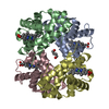
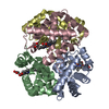
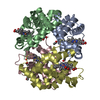
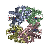
| Title | X-ray structure of an intermediate along the oxidation pathway of Trematomus bernacchii hemoglobin | ||||||
 Components Components |
| ||||||
 Keywords Keywords | OXYGEN TRANSPORT / hemoglobin / intermediate quaternary structure / Acetylation / Heme / Iron / Metal-binding / Transport | ||||||
| Function / homology |  Function and homology information Function and homology informationhaptoglobin binding / organic acid binding / haptoglobin-hemoglobin complex / hemoglobin complex / oxygen transport / hydrogen peroxide catabolic process / oxygen carrier activity / peroxidase activity / oxygen binding / blood microparticle ...haptoglobin binding / organic acid binding / haptoglobin-hemoglobin complex / hemoglobin complex / oxygen transport / hydrogen peroxide catabolic process / oxygen carrier activity / peroxidase activity / oxygen binding / blood microparticle / iron ion binding / heme binding / metal ion binding Similarity search - Function | ||||||
| Biological species |  Trematomus bernacchii (emerald rockcod) Trematomus bernacchii (emerald rockcod) | ||||||
| Method |  X-RAY DIFFRACTION / SYNCHROTRON / MOLECULAR REPLACEMENT / Resolution: 1.4 Å X-RAY DIFFRACTION / SYNCHROTRON / MOLECULAR REPLACEMENT / Resolution: 1.4 Å | ||||||
 Authors Authors | Merlino, A. / Vitagliano, L. / Sica, F. / Vergara, A. / Mazzarella, L. | ||||||
 Citation Citation | Journal: Biopolymers / Year: 2009 Title: Combined crystallographic and spectroscopic analysis of Trematomus bernacchii hemoglobin highlights analogies and differences in the peculiar oxidation pathway of Antarctic fish hemoglobins Authors: Merlino, A. / Vitagliano, L. / Howes, B.D. / Verde, C. / di Prisco, G. / Smulevich, G. / Sica, F. / Vergara, A. #1: Journal: J.Am.Chem.Soc. / Year: 2008Title: Spectroscopic and crystallographic characterization of a tetrameric hemoglobin oxidation reveals structural features of the functional intermediate relaxed/tense state Authors: Vitagliano, L. / Vergara, A. / Bonomi, G. / Merlino, A. / Verde, C. / di Prisco, G. / Howes, B.D. / Smulevich, G. / Mazzarella, L. | ||||||
| History |
|
- Structure visualization
| Structure viewer | Molecule: MolmilJmol/JSmol |
|---|
- Downloads & links
-Download
| PDBx/mmCIF format | 3gkv.cif.gz | 78.8 KB | Display | PDBx/mmCIF format |
|---|---|---|---|---|
| PDB format | pdb3gkv.ent.gz | 57.5 KB | Display | PDB format |
| PDBx/mmJSON format | 3gkv.json.gz | Tree view | PDBx/mmJSON format | |
| Others |  Other downloads Other downloads |
-Validation report
| Arichive directory | https://data.pdbj.org/pub/pdb/validation_reports/gk/3gkvftp://data.pdbj.org/pub/pdb/validation_reports/gk/3gkv | HTTPS FTP |
|---|
-Related structure data
| Related structure data |  2pegS S: Starting model for refinement |
|---|---|
| Similar structure data |


-Links
 PDBj
PDBj


- Assembly
| Deposited unit | 
| ||||||||
|---|---|---|---|---|---|---|---|---|---|
| 1 | 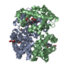
| ||||||||
| Unit cell |
|
-Components
| #1: Protein | Mass: 15683.271 Da / Num. of mol.: 1 / Source method: isolated from a natural source / Source: (natural) Trematomus bernacchii (emerald rockcod) / References: UniProt: P80043 | ||||||
|---|---|---|---|---|---|---|---|
| #2: Protein | Mass: 16153.368 Da / Num. of mol.: 1 / Source method: isolated from a natural source / Source: (natural) Trematomus bernacchii (emerald rockcod) / References: UniProt: P80044 | ||||||
| #3: Chemical |   Mass: 616.487 Da / Num. of mol.: 2 / Source method: obtained synthetically / Formula: C34H32FeN4O4 Mass: 616.487 Da / Num. of mol.: 2 / Source method: obtained synthetically / Formula: C34H32FeN4O4#4: Chemical |   Mass: 28.010 Da / Num. of mol.: 2 / Source method: obtained synthetically / Formula: CO Mass: 28.010 Da / Num. of mol.: 2 / Source method: obtained synthetically / Formula: CO#5: Water | ChemComp-HOH / |  Mass: 18.015 Da / Num. of mol.: 241 / Source method: isolated from a natural source / Formula: H2O Mass: 18.015 Da / Num. of mol.: 241 / Source method: isolated from a natural source / Formula: H2OHas protein modification | Y | |
-Experimental details
-Experiment
| Experiment | Method: X-RAY DIFFRACTION / Number of used crystals: 1 |
|---|
- Sample preparation
| Crystal | Density Matthews: 3.26 Å3/Da / Density % sol: 62.24 % |
|---|---|
| Crystal grow | Temperature: 298 K / Method: liquid diffusion / pH: 7.6 Details: protein concentration of 6mg/ml, 14% Mpeg 5000 in 50mM Tris/HCl buffer ph 7.6, LIQUID DIFFUSION, temperature 298K |
-Data collection
| Diffraction | Mean temperature: 100 K |
|---|---|
| Diffraction source | Source: SYNCHROTRON / Site: ESRF  / Beamline: ID14-1 / Wavelength: 1 Å / Beamline: ID14-1 / Wavelength: 1 Å |
| Detector | Type: ADSC QUANTUM 210 / Detector: CCD / Date: Mar 19, 2000 |
| Radiation | Protocol: SINGLE WAVELENGTH / Monochromatic (M) / Laue (L): M / Scattering type: x-ray |
| Radiation wavelength | Wavelength: 1 Å / Relative weight: 1 |
| Reflection | Resolution: 1.4→30 Å / Num. obs: 80090 / % possible obs: 99.9 % / Rmerge(I) obs: 0.038 |
- Processing
| Software |
| ||||||||||||||||||||
|---|---|---|---|---|---|---|---|---|---|---|---|---|---|---|---|---|---|---|---|---|---|
| Refinement | Method to determine structure: MOLECULAR REPLACEMENT Starting model: PDB ENTRY 2PEG Resolution: 1.4→28.25 Å / Isotropic thermal model: Isotropic / Cross valid method: THROUGHOUT / σ(F): 0 / σ(I): 0 / Stereochemistry target values: Engh & Huber
| ||||||||||||||||||||
| Refinement step | Cycle: LAST / Resolution: 1.4→28.25 Å
|
