 Movie
Movie Controller

[English] 日本語
 Yorodumi
- PDB-4erl: Crystal structure of the lysine riboswitch bound to a lysine-glyc... -
+ Open data

- Basic information
| Entry | Database: PDB / ID: 4erl | ||||||
|---|---|---|---|---|---|---|---|
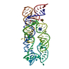
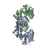
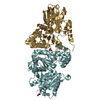
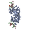
| Title | Crystal structure of the lysine riboswitch bound to a lysine-glycine dipeptide | ||||||
 Components Components | Lysine riboswitch RNA | ||||||
 Keywords Keywords | TRANSCRIPTION / Riboswitch aptamer domain / regulatory mRNA | ||||||
| Function / homology | GLYCINE / LYSINE / RNA / RNA (> 10) / RNA (> 100) Function and homology information Function and homology information | ||||||
| Biological species |   Thermotoga Maritima (bacteria) Thermotoga Maritima (bacteria) | ||||||
| Method |  X-RAY DIFFRACTION / MOLECULAR REPLACEMENT / Resolution: 3 Å X-RAY DIFFRACTION / MOLECULAR REPLACEMENT / Resolution: 3 Å | ||||||
 Authors Authors | Garst, A.D. / Porter, E. / Batey, R.T. | ||||||
 Citation Citation | Journal: J.Mol.Biol. / Year: 2012 Title: Insights into the regulatory landscape of the lysine riboswitch. Authors: Garst, A.D. / Porter, E.B. / Batey, R.T. | ||||||
| History |
|
- Structure visualization
| Structure viewer | Molecule: MolmilJmol/JSmol |
|---|
- Downloads & links
-Download
| PDBx/mmCIF format | 4erl.cif.gz | 181.2 KB | Display | PDBx/mmCIF format |
|---|---|---|---|---|
| PDB format | pdb4erl.ent.gz | 142.9 KB | Display | PDB format |
| PDBx/mmJSON format | 4erl.json.gz | Tree view | PDBx/mmJSON format | |
| Others |  Other downloads Other downloads |
-Validation report
| Arichive directory | https://data.pdbj.org/pub/pdb/validation_reports/er/4erlftp://data.pdbj.org/pub/pdb/validation_reports/er/4erl | HTTPS FTP |
|---|
-Related structure data
| Related structure data |  4erjC  3d0uS S: Starting model for refinement C: citing same article ( |
|---|---|
| Similar structure data |


-Links
 PDBj
PDBj


- Assembly
| Deposited unit | 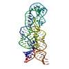
| ||||||||
|---|---|---|---|---|---|---|---|---|---|
| 1 |
| ||||||||
| Unit cell |
|
-Components
| #1: RNA chain | Mass: 52465.289 Da / Num. of mol.: 1 / Fragment: Lysine riboswitch aptamer domain Source method: isolated from a genetically manipulated source Source: (gene. exp.) Thermotoga Maritima (bacteria) / Gene: asd |
|---|---|
| #2: Chemical | ChemComp-LYS /   Type: L-peptide linking / Mass: 147.195 Da / Num. of mol.: 1 / Source method: obtained synthetically / Formula: C6H15N2O2 Type: L-peptide linking / Mass: 147.195 Da / Num. of mol.: 1 / Source method: obtained synthetically / Formula: C6H15N2O2 |
| #3: Chemical | ChemComp-GLY /   Type: peptide linking / Mass: 75.067 Da / Num. of mol.: 1 / Source method: obtained synthetically / Formula: C2H5NO2 Type: peptide linking / Mass: 75.067 Da / Num. of mol.: 1 / Source method: obtained synthetically / Formula: C2H5NO2 |
| #4: Water | ChemComp-HOH /  Mass: 18.015 Da / Num. of mol.: 10 / Source method: isolated from a natural source / Formula: H2O Mass: 18.015 Da / Num. of mol.: 10 / Source method: isolated from a natural source / Formula: H2O |
-Experimental details
-Experiment
| Experiment | Method: X-RAY DIFFRACTION / Number of used crystals: 1 |
|---|
- Sample preparation
| Crystal | Density Matthews: 4.64 Å3/Da / Density % sol: 73.52 % |
|---|---|
| Crystal grow | Temperature: 303 K / Method: vapor diffusion, hanging drop / pH: 7 Details: 10 mM Na-HEPES pH 7.0, 2 M Li2SO4, and 5 mM MgCl2, VAPOR DIFFUSION, HANGING DROP, temperature 303K |
-Data collection
| Diffraction | Mean temperature: 80 K |
|---|---|
| Diffraction source | Source: ROTATING ANODE / Type: RIGAKU RU300 / Wavelength: 1.5418 Å |
| Detector | Type: ADSC QUANTUM 315 / Detector: CCD / Date: Apr 12, 2010 |
| Radiation | Protocol: SINGLE WAVELENGTH / Monochromatic (M) / Laue (L): M / Scattering type: x-ray |
| Radiation wavelength | Wavelength: 1.5418 Å / Relative weight: 1 |
| Reflection | Resolution: 3→19.62 Å / Num. all: 18847 / % possible obs: 97.8 % / Observed criterion σ(F): 1.97 / Observed criterion σ(I): 2 / Redundancy: 1.92 % / Biso Wilson estimate: 47.24 Å2 / Rmerge(I) obs: 0.113 / Rsym value: 0.113 / Net I/σ(I): 5.1 |
| Reflection shell | Resolution: 3→3.11 Å / Redundancy: 1.93 % / Rmerge(I) obs: 0.321 / Mean I/σ(I) obs: 1.9 / Num. unique all: 1853 / Rsym value: 0.321 / % possible all: 96.5 |
- Processing
| Software |
| ||||||||||||||||||||||||||||||||||||||||||||||||||||||||
|---|---|---|---|---|---|---|---|---|---|---|---|---|---|---|---|---|---|---|---|---|---|---|---|---|---|---|---|---|---|---|---|---|---|---|---|---|---|---|---|---|---|---|---|---|---|---|---|---|---|---|---|---|---|---|---|---|---|
| Refinement | Method to determine structure: MOLECULAR REPLACEMENT Starting model: PDB ENTRY 3D0U) Resolution: 3→19.62 Å / SU ML: 0.43 / σ(F): 1.97 / Phase error: 26.54 / Stereochemistry target values: ML
| ||||||||||||||||||||||||||||||||||||||||||||||||||||||||
| Solvent computation | Shrinkage radii: 0.9 Å / VDW probe radii: 1.11 Å / Solvent model: FLAT BULK SOLVENT MODEL / Bsol: 11.901 Å2 / ksol: 0.254 e/Å3 | ||||||||||||||||||||||||||||||||||||||||||||||||||||||||
| Displacement parameters |
| ||||||||||||||||||||||||||||||||||||||||||||||||||||||||
| Refine analyze | Luzzati d res low obs: 0.43 Å | ||||||||||||||||||||||||||||||||||||||||||||||||||||||||
| Refinement step | Cycle: LAST / Resolution: 3→19.62 Å
| ||||||||||||||||||||||||||||||||||||||||||||||||||||||||
| Refine LS restraints |
| ||||||||||||||||||||||||||||||||||||||||||||||||||||||||
| LS refinement shell |
| ||||||||||||||||||||||||||||||||||||||||||||||||||||||||
| Refinement TLS params. | Method: refined / Origin x: -23.1393 Å / Origin y: 45.4269 Å / Origin z: 1264.8736 Å
| ||||||||||||||||||||||||||||||||||||||||||||||||||||||||
| Refinement TLS group | Selection details: all |
