 Movie
Movie Controller


+ Open data

- Basic information
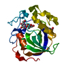
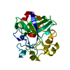
| Entry | Database: PDB / ID: 4eng | |||||||||
|---|---|---|---|---|---|---|---|---|---|---|
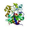
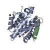
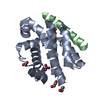
| Title | STRUCTURE OF ENDOGLUCANASE V CELLOHEXAOSE COMPLEX | |||||||||
 Components Components | ENDOGLUCANASE V CELLOHEXAOSE COMPLEX | |||||||||
 Keywords Keywords | GLYCOSYL HYDROLASE / HYDROLASE / ENDOGLUCANASE | |||||||||
| Function / homology |  Function and homology information Function and homology information | |||||||||
| Biological species |  Humicola insolens (fungus) Humicola insolens (fungus) | |||||||||
| Method |  X-RAY DIFFRACTION / MOLECULAR REPLACEMENT / Resolution: 1.9 Å X-RAY DIFFRACTION / MOLECULAR REPLACEMENT / Resolution: 1.9 Å | |||||||||
 Authors Authors | Davies, G.J. / Schulein, M. | |||||||||
 Citation Citation | Journal: Acta Crystallogr.,Sect.D / Year: 1996 Title: Structure determination and refinement of the Humicola insolens endoglucanase V at 1.5 A resolution. Authors: Davies, G.J. / Dodson, G. / Moore, M.H. / Tolley, S.P. / Dauter, Z. / Wilson, K.S. / Rasmussen, G. / Schulein, M. #1: Journal: Biochemistry / Year: 1995Title: Structures of Oligosaccharide-Bound Forms of the Endoglucanase V from Humicola Insolens at 1.9 A Resolution Authors: Davies, G.J. / Tolley, S.P. / Henrissat, B. / Hjort, C. / Schulein, M. #2: Journal: Nature / Year: 1993Title: Structure and Function of Endoglucanase V Authors: Davies, G.J. / Dodson, G.G. / Hubbard, R.E. / Tolley, S.P. / Dauter, Z. / Wilson, K.S. / Hjort, C. / Mikkelsen, J.M. / Rasmussen, G. / Schulein, M. | |||||||||
| History |
|
- Structure visualization
| Structure viewer | Molecule: MolmilJmol/JSmol |
|---|
- Downloads & links
-Download
| PDBx/mmCIF format | 4eng.cif.gz | 58.1 KB | Display | PDBx/mmCIF format |
|---|---|---|---|---|
| PDB format | pdb4eng.ent.gz | 40.7 KB | Display | PDB format |
| PDBx/mmJSON format | 4eng.json.gz | Tree view | PDBx/mmJSON format | |
| Others |  Other downloads Other downloads |
-Validation report
| Arichive directory | https://data.pdbj.org/pub/pdb/validation_reports/en/4engftp://data.pdbj.org/pub/pdb/validation_reports/en/4eng | HTTPS FTP |
|---|
-Related structure data
| Related structure data | 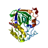 3engC 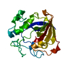 2engS S: Starting model for refinement C: citing same article ( |
|---|---|
| Similar structure data |


-Links
 PDBj
PDBj- Assembly
| Deposited unit | 
| ||||||||
|---|---|---|---|---|---|---|---|---|---|
| 1 |
| ||||||||
| Unit cell |
|
-Components
| #1: Protein | Mass: 22544.984 Da / Num. of mol.: 1 / Fragment: CATALYTIC CORE, RESIDUES 1 - 210 / Mutation: D10N Source method: isolated from a genetically manipulated source Source: (gene. exp.) Humicola insolens (fungus) / References: UniProt: P43316, cellulase | ||||||
|---|---|---|---|---|---|---|---|
| #2: Polysaccharide | 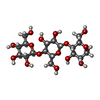  Source method: isolated from a genetically manipulated source Details: oligosaccharide / References: alpha-cellotriose #3: Water | ChemComp-HOH / |  Mass: 18.015 Da / Num. of mol.: 132 / Source method: isolated from a natural source / Formula: H2O Mass: 18.015 Da / Num. of mol.: 132 / Source method: isolated from a natural source / Formula: H2OCompound details | THIS IS AN INACTIVE MUTANT (D10N) COMPLEX WITH THE SUBSTRATE CELLOHEXAOSE. TWO HALF CELLOHEXAOSES ...THIS IS AN INACTIVE MUTANT (D10N) COMPLEX WITH THE SUBSTRATE CELLOHEXAO | Has protein modification | Y | |
-Experimental details
-Experiment
| Experiment | Method: X-RAY DIFFRACTION / Number of used crystals: 1 |
|---|
- Sample preparation
| Crystal | Density Matthews: 2.01 Å3/Da / Density % sol: 38.83 % | ||||||||||||||||||||
|---|---|---|---|---|---|---|---|---|---|---|---|---|---|---|---|---|---|---|---|---|---|
| Crystal grow | pH: 8 Details: 10MG/ML ENZYME IN 20MM TRIS-HCL BUFFER PH 8.0. PRECIPITANT 18%(W/V) PEG 8K. CO-CRYSTALLISED WITH 5MM CELLOHEXAOSE | ||||||||||||||||||||
| Crystal grow | *PLUS Method: vapor diffusion, hanging drop / Details: Davies, G.J., (1993) Nature, 365, 362. | ||||||||||||||||||||
| Components of the solutions | *PLUS
|
-Data collection
| Diffraction | Mean temperature: 293 K |
|---|---|
| Diffraction source | Source: ROTATING ANODE / Type: RIGAKU RUH2R / Wavelength: 1.5418 |
| Detector | Type: RIGAKU / Detector: IMAGE PLATE / Date: 1993 |
| Radiation | Monochromator: GRAPHITE(002) / Monochromatic (M) / Laue (L): M / Scattering type: x-ray |
| Radiation wavelength | Wavelength: 1.5418 Å / Relative weight: 1 |
| Reflection | Resolution: 1.9→20 Å / Num. obs: 13502 / % possible obs: 95.1 % / Observed criterion σ(I): -999 / Redundancy: 3.2 % / Rmerge(I) obs: 0.085 / Net I/σ(I): 12.56 |
| Reflection shell | Resolution: 1.9→2 Å / Redundancy: 1.4 % / Rmerge(I) obs: 0.157 / Mean I/σ(I) obs: 4.48 / % possible all: 64 |
- Processing
| Software |
| ||||||||||||||||||||||||||||||||||||||||||||||||||||||||||||||||||||||||||||||||||||
|---|---|---|---|---|---|---|---|---|---|---|---|---|---|---|---|---|---|---|---|---|---|---|---|---|---|---|---|---|---|---|---|---|---|---|---|---|---|---|---|---|---|---|---|---|---|---|---|---|---|---|---|---|---|---|---|---|---|---|---|---|---|---|---|---|---|---|---|---|---|---|---|---|---|---|---|---|---|---|---|---|---|---|---|---|---|
| Refinement | Method to determine structure: MOLECULAR REPLACEMENT Starting model: PDB ENTRY 2ENG Resolution: 1.9→10 Å /
| ||||||||||||||||||||||||||||||||||||||||||||||||||||||||||||||||||||||||||||||||||||
| Refinement step | Cycle: LAST / Resolution: 1.9→10 Å
| ||||||||||||||||||||||||||||||||||||||||||||||||||||||||||||||||||||||||||||||||||||
| Refine LS restraints |
|
