 Movie
Movie Controller

[English] 日本語
 Yorodumi
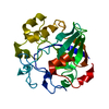
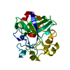
- PDB-1oa7: Structure of Melanocarpus albomyces endoglucanase in complex with... -
+ Open data

- Basic information
| Entry | Database: PDB / ID: 1oa7 | |||||||||
|---|---|---|---|---|---|---|---|---|---|---|
| Title | Structure of Melanocarpus albomyces endoglucanase in complex with cellobiose | |||||||||
 Components Components | CELLULASE | |||||||||
 Keywords Keywords | HYDROLASE / CELLULASE / CELLULOSE DEGRADATION / GLYCOSIDE HYDROLASES | |||||||||
| Function / homology |  Function and homology information Function and homology information | |||||||||
| Biological species |  MELANOCARPUS ALBOMYCES (fungus) MELANOCARPUS ALBOMYCES (fungus) | |||||||||
| Method |  X-RAY DIFFRACTION / MOLECULAR REPLACEMENT / Resolution: 2 Å X-RAY DIFFRACTION / MOLECULAR REPLACEMENT / Resolution: 2 Å | |||||||||
 Authors Authors | Hirvonen, M. / Papageorgiou, A.C. | |||||||||
 Citation Citation | Journal: J.Mol.Biol. / Year: 2003 Title: Crystal Structure of a Family 45 Endoglucanase from Melanocarpus Albomyces: Mechanistic Implications Based on the Free and Cellobiose-Bound Forms Authors: Hirvonen, M. / Papageorgiou, A.C. | |||||||||
| History |
| |||||||||
| Remark 700 | SHEET DETERMINATION METHOD: DSSP THE SHEETS PRESENTED AS "AA" IN EACH CHAIN ON SHEET RECORDS BELOW ... SHEET DETERMINATION METHOD: DSSP THE SHEETS PRESENTED AS "AA" IN EACH CHAIN ON SHEET RECORDS BELOW IS ACTUALLY AN 6-STRANDED BARREL THIS IS REPRESENTED BY A 7-STRANDED SHEET IN WHICH THE FIRST AND LAST STRANDS ARE IDENTICAL. |
- Structure visualization
| Structure viewer | Molecule: MolmilJmol/JSmol |
|---|
- Downloads & links
-Download
| PDBx/mmCIF format | 1oa7.cif.gz | 56.8 KB | Display | PDBx/mmCIF format |
|---|---|---|---|---|
| PDB format | pdb1oa7.ent.gz | 40.5 KB | Display | PDB format |
| PDBx/mmJSON format | 1oa7.json.gz | Tree view | PDBx/mmJSON format | |
| Others |  Other downloads Other downloads |
-Validation report
| Arichive directory | https://data.pdbj.org/pub/pdb/validation_reports/oa/1oa7ftp://data.pdbj.org/pub/pdb/validation_reports/oa/1oa7 | HTTPS FTP |
|---|
-Related structure data
| Related structure data |  1oa9C 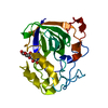 3engS C: citing same article ( S: Starting model for refinement |
|---|---|
| Similar structure data |


-Links
 PDBj
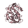
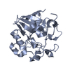
PDBj- Assembly
| Deposited unit | 
| ||||||||
|---|---|---|---|---|---|---|---|---|---|
| 1 |
| ||||||||
| Unit cell |
|
-Components
| #1: Protein | Mass: 22932.221 Da / Num. of mol.: 1 / Source method: isolated from a natural source / Source: (natural) MELANOCARPUS ALBOMYCES (fungus) / References: UniProt: Q8J0K8, cellulase |
|---|---|
| #2: Polysaccharide | beta-D-glucopyranose-(1-4)-beta-D-glucopyranose / beta-cellobiose  Source method: isolated from a genetically manipulated source Details: oligosaccharide / References: beta-cellobiose |
| #3: Water | ChemComp-HOH /  Mass: 18.015 Da / Num. of mol.: 223 / Source method: isolated from a natural source / Formula: H2O Mass: 18.015 Da / Num. of mol.: 223 / Source method: isolated from a natural source / Formula: H2O |
| Has protein modification | Y |
-Experimental details
-Experiment
| Experiment | Method: X-RAY DIFFRACTION / Number of used crystals: 1 |
|---|
- Sample preparation
| Crystal | Density Matthews: 2.5 Å3/Da / Density % sol: 50 % |
|---|---|
| Crystal grow | pH: 4.6 / Details: PEG 4000 15% W/V, 0.15 M KOAC, 0.1 M NAOAC PH 4.6 |
| Crystal grow | *PLUS Method: unknown / Details: Hirvonen, M., (2002) Acta Cryst., D58, 336. |
-Data collection
| Diffraction | Mean temperature: 100 K |
|---|---|
| Diffraction source | Source: ROTATING ANODE / Type: RIGAKU RU200 / Wavelength: 1.5418 |
| Detector | Type: MARRESEARCH / Detector: IMAGE PLATE / Date: Jul 15, 2001 / Details: OSMIC |
| Radiation | Protocol: SINGLE WAVELENGTH / Monochromatic (M) / Laue (L): M / Scattering type: x-ray |
| Radiation wavelength | Wavelength: 1.5418 Å / Relative weight: 1 |
| Reflection | Resolution: 2→50 Å / Num. obs: 133688 / % possible obs: 92.4 % / Observed criterion σ(I): -3 / Redundancy: 9.9 % / Biso Wilson estimate: 16.8 Å2 / Rmerge(I) obs: 0.066 / Net I/σ(I): 12.1 |
| Reflection shell | Resolution: 2→2.06 Å / Rmerge(I) obs: 0.408 / Mean I/σ(I) obs: 3.6 / % possible all: 89.1 |
| Reflection | *PLUS Highest resolution: 2 Å / Num. obs: 13447 / Num. measured all: 133688 / Rmerge(I) obs: 0.084 |
| Reflection shell | *PLUS % possible obs: 89.1 % |
- Processing
| Software |
| ||||||||||||||||||||||||||||||||||||||||||||||||||||||||||||||||||||||||||||||||
|---|---|---|---|---|---|---|---|---|---|---|---|---|---|---|---|---|---|---|---|---|---|---|---|---|---|---|---|---|---|---|---|---|---|---|---|---|---|---|---|---|---|---|---|---|---|---|---|---|---|---|---|---|---|---|---|---|---|---|---|---|---|---|---|---|---|---|---|---|---|---|---|---|---|---|---|---|---|---|---|---|---|
| Refinement | Method to determine structure: MOLECULAR REPLACEMENT Starting model: PDB ENTRY 3ENG Resolution: 2→50 Å / Rfactor Rfree error: 0.01 / Data cutoff high absF: 1500347 / Isotropic thermal model: RESTRAINED / Cross valid method: THROUGHOUT / σ(F): 0
| ||||||||||||||||||||||||||||||||||||||||||||||||||||||||||||||||||||||||||||||||
| Solvent computation | Solvent model: FLAT / Bsol: 49.5 Å2 / ksol: 0.36 e/Å3 | ||||||||||||||||||||||||||||||||||||||||||||||||||||||||||||||||||||||||||||||||
| Displacement parameters | Biso mean: 20.9 Å2 | ||||||||||||||||||||||||||||||||||||||||||||||||||||||||||||||||||||||||||||||||
| Refine analyze |
| ||||||||||||||||||||||||||||||||||||||||||||||||||||||||||||||||||||||||||||||||
| Refinement step | Cycle: LAST / Resolution: 2→50 Å
| ||||||||||||||||||||||||||||||||||||||||||||||||||||||||||||||||||||||||||||||||
| Refine LS restraints |
| ||||||||||||||||||||||||||||||||||||||||||||||||||||||||||||||||||||||||||||||||
| LS refinement shell | Resolution: 2→2.13 Å / Rfactor Rfree error: 0.026 / Total num. of bins used: 6
| ||||||||||||||||||||||||||||||||||||||||||||||||||||||||||||||||||||||||||||||||
| Xplor file |
| ||||||||||||||||||||||||||||||||||||||||||||||||||||||||||||||||||||||||||||||||
| Refinement | *PLUS Lowest resolution: 50 Å / % reflection Rfree: 5 % | ||||||||||||||||||||||||||||||||||||||||||||||||||||||||||||||||||||||||||||||||
| Solvent computation | *PLUS | ||||||||||||||||||||||||||||||||||||||||||||||||||||||||||||||||||||||||||||||||
| Displacement parameters | *PLUS | ||||||||||||||||||||||||||||||||||||||||||||||||||||||||||||||||||||||||||||||||
| Refine LS restraints | *PLUS
|
