 Movie
Movie Controller

[English] 日本語
 Yorodumi
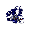
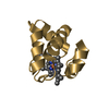
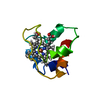
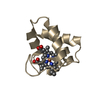
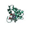
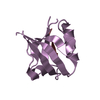
- PDB-4eic: Crystal structure of reduced cytochrome c6 from Synechococcus sp.... -
+ Open data

- Basic information
| Entry | Database: PDB / ID: 4eic | |||||||||
|---|---|---|---|---|---|---|---|---|---|---|
| Title | Crystal structure of reduced cytochrome c6 from Synechococcus sp. PCC 7002 at ultra-high resolution | |||||||||
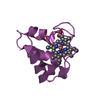
 Components Components | Cytochrome c6 | |||||||||
 Keywords Keywords | ELECTRON TRANSPORT / Cytochrome C6 | |||||||||
| Function / homology |  Function and homology information Function and homology informationplasma membrane-derived thylakoid lumen / photosynthesis / electron transfer activity / iron ion binding / heme binding Similarity search - Function | |||||||||
| Biological species |  Synechococcus sp. (bacteria) Synechococcus sp. (bacteria) | |||||||||
| Method |  X-RAY DIFFRACTION / SYNCHROTRON / MOLECULAR REPLACEMENT / Resolution: 0.84 Å X-RAY DIFFRACTION / SYNCHROTRON / MOLECULAR REPLACEMENT / Resolution: 0.84 Å | |||||||||
 Authors Authors | Krzywda, S. / Bialek, W. / Jaskolski, M. / Szczepaniak, A. | |||||||||
 Citation Citation | Journal: Acta Crystallogr.,Sect.D / Year: 2014 Title: Insights into the relationship between the haem-binding pocket and the redox potential of c6 cytochromes: four atomic resolution structures of c6 and c6-like proteins from Synechococcus sp. PCC 7002. Authors: Bialek, W. / Krzywda, S. / Zatwarnicki, P. / Jaskolski, M. / Kolesinski, P. / Szczepaniak, A. #1: Journal: Febs J. / Year: 2009Title: Atomic-resolution structure of reduced cyanobacterial cytochrome c6 with an unusual sequence insertion. Authors: Bialek, W. / Krzywda, S. / Jaskolski, M. / Szczepaniak, A. #2: Journal: Biochemistry / Year: 2008 Title: Deeply branching c6-like cytochromes of cyanobacteria. Authors: Bialek, W. / Nelson, M. / Tamiola, K. / Kallas, T. / Szczepaniak, A. | |||||||||
| History |
|
- Structure visualization
| Structure viewer | Molecule: MolmilJmol/JSmol |
|---|
- Downloads & links
-Download
| PDBx/mmCIF format | 4eic.cif.gz | 56.6 KB | Display | PDBx/mmCIF format |
|---|---|---|---|---|
| PDB format | pdb4eic.ent.gz | 39 KB | Display | PDB format |
| PDBx/mmJSON format | 4eic.json.gz | Tree view | PDBx/mmJSON format | |
| Others |  Other downloads Other downloads |
-Validation report
| Arichive directory | https://data.pdbj.org/pub/pdb/validation_reports/ei/4eicftp://data.pdbj.org/pub/pdb/validation_reports/ei/4eic | HTTPS FTP |
|---|
-Related structure data
| Related structure data | 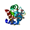 4eidC 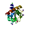 4eieC 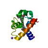 4eifC  3dr0S S: Starting model for refinement C: citing same article ( |
|---|---|
| Similar structure data |


-Links
 PDBj
PDBj


- Assembly
| Deposited unit | 
| ||||||||
|---|---|---|---|---|---|---|---|---|---|
| 1 |
| ||||||||
| Unit cell |
|
-Components
| #1: Protein | Mass: 9446.432 Da / Num. of mol.: 1 / Fragment: UNP Residues 25-117 Source method: isolated from a genetically manipulated source Source: (gene. exp.) Synechococcus sp. (bacteria) / Strain: ATCC 27264 / PCC 7002 / PR-6 / Description: co-expression with pEC86 / Gene: petJ, petJ1, SYNPCC7002_A0167 / Plasmid: pUCJ1 / Production host: |
|---|---|
| #2: Chemical | ChemComp-HEC /   Mass: 618.503 Da / Num. of mol.: 1 / Source method: obtained synthetically / Formula: C34H34FeN4O4 Mass: 618.503 Da / Num. of mol.: 1 / Source method: obtained synthetically / Formula: C34H34FeN4O4 |
| #3: Chemical | ChemComp-MES /   Mass: 195.237 Da / Num. of mol.: 1 / Source method: obtained synthetically / Formula: C6H13NO4S / Comment: pH buffer*YM Mass: 195.237 Da / Num. of mol.: 1 / Source method: obtained synthetically / Formula: C6H13NO4S / Comment: pH buffer*YM |
| #4: Water | ChemComp-HOH /  Mass: 18.015 Da / Num. of mol.: 120 / Source method: isolated from a natural source / Formula: H2O Mass: 18.015 Da / Num. of mol.: 120 / Source method: isolated from a natural source / Formula: H2O |
| Has protein modification | Y |
-Experimental details
-Experiment
| Experiment | Method: X-RAY DIFFRACTION / Number of used crystals: 1 |
|---|
- Sample preparation
| Crystal | Density Matthews: 2.02 Å3/Da / Density % sol: 39.09 % |
|---|---|
| Crystal grow | Temperature: 292 K / Method: vapor diffusion, hanging drop / pH: 6.5 Details: 2.2M Ammonium sulphate, 0.1M MES, pH 6.5, VAPOR DIFFUSION, HANGING DROP, temperature 292K |
-Data collection
| Diffraction | Mean temperature: 100 K |
|---|---|
| Diffraction source | Source: SYNCHROTRON / Site: EMBL/DESY, HAMBURG  / Beamline: X11 / Wavelength: 0.8166 Å / Beamline: X11 / Wavelength: 0.8166 Å |
| Detector | Type: MAR CCD 165 mm / Detector: CCD / Date: Dec 14, 2006 / Details: mirrors |
| Radiation | Monochromator: Ge / Protocol: SINGLE WAVELENGTH / Monochromatic (M) / Laue (L): M / Scattering type: x-ray |
| Radiation wavelength | Wavelength: 0.8166 Å / Relative weight: 1 |
| Reflection | Resolution: 0.84→32.55 Å / Num. all: 64908 / Num. obs: 64908 / % possible obs: 94.8 % / Observed criterion σ(F): 0 / Observed criterion σ(I): -3 / Redundancy: 6 % / Biso Wilson estimate: 9.45 Å2 / Rmerge(I) obs: 0.063 / Net I/σ(I): 15.21 |
| Reflection shell | Resolution: 0.84→0.86 Å / Redundancy: 4.3 % / Rmerge(I) obs: 0.507 / Mean I/σ(I) obs: 3.23 / Num. unique all: 4069 / % possible all: 80.6 |
- Processing
| Software |
| |||||||||||||||||||||||||||||||||
|---|---|---|---|---|---|---|---|---|---|---|---|---|---|---|---|---|---|---|---|---|---|---|---|---|---|---|---|---|---|---|---|---|---|---|
| Refinement | Method to determine structure: MOLECULAR REPLACEMENT Starting model: PDB ENTRY 3DR0 Resolution: 0.84→32.55 Å / Num. parameters: 7849 / Num. restraintsaints: 7790 / Cross valid method: FREE R / σ(F): 0 / Stereochemistry target values: Engh & Huber Details: ANISOTROPIC REFINEMENT, HYDROGENS HAVE BEEN ADDED IN THE RIDING POSITIONS
| |||||||||||||||||||||||||||||||||
| Refine analyze | Num. disordered residues: 12 / Occupancy sum hydrogen: 664.26 / Occupancy sum non hydrogen: 800.71 | |||||||||||||||||||||||||||||||||
| Refinement step | Cycle: LAST / Resolution: 0.84→32.55 Å
| |||||||||||||||||||||||||||||||||
| Refine LS restraints |
|
