 Movie
Movie Controller

[English] 日本語
 Yorodumi
- PDB-4eia: Activator of the 2-Hydroxyisocaproyl-CoA Dehydratase from Clostri... -
+ Open data

- Basic information
| Entry | Database: PDB / ID: 4eia | ||||||
|---|---|---|---|---|---|---|---|
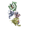
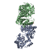
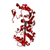
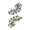
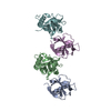
| Title | Activator of the 2-Hydroxyisocaproyl-CoA Dehydratase from Clostridium difficile without nucleotide | ||||||
 Components Components | Activator of 2-hydroxyisocaproyl-CoA dehydratase | ||||||
 Keywords Keywords | ELECTRON TRANSPORT / actin fold / ATPase / electron transfer / ATP/ADP binding / 2-hydroxyisocaproyl-CoA dehydratase binding | ||||||
| Function / homology |  Function and homology information Function and homology informationL-leucine metabolic process / Hydrolases / 4 iron, 4 sulfur cluster binding / hydrolase activity / ATP binding / metal ion binding Similarity search - Function | ||||||
| Biological species |  Clostridium difficile (bacteria) Clostridium difficile (bacteria) | ||||||
| Method |  X-RAY DIFFRACTION / SYNCHROTRON / MOLECULAR REPLACEMENT / Resolution: 3 Å X-RAY DIFFRACTION / SYNCHROTRON / MOLECULAR REPLACEMENT / Resolution: 3 Å | ||||||
 Authors Authors | Knauer, S.H. / Dobbek, H. | ||||||
 Citation Citation | Journal: Biochemistry / Year: 2012 Title: On the ATP-Dependent Activation of the Radical Enzyme (R)-2-Hydroxyisocaproyl-CoA Dehydratase. Authors: Knauer, S.H. / Buckel, W. / Dobbek, H. | ||||||
| History |
|
- Structure visualization
| Structure viewer | Molecule: MolmilJmol/JSmol |
|---|
- Downloads & links
-Download
| PDBx/mmCIF format | 4eia.cif.gz | 112.4 KB | Display | PDBx/mmCIF format |
|---|---|---|---|---|
| PDB format | pdb4eia.ent.gz | 87.1 KB | Display | PDB format |
| PDBx/mmJSON format | 4eia.json.gz | Tree view | PDBx/mmJSON format | |
| Others |  Other downloads Other downloads |
-Validation report
| Arichive directory | https://data.pdbj.org/pub/pdb/validation_reports/ei/4eiaftp://data.pdbj.org/pub/pdb/validation_reports/ei/4eia | HTTPS FTP |
|---|
-Related structure data
| Related structure data |  4ehtSC  4ehuC S: Starting model for refinement C: citing same article ( |
|---|---|
| Similar structure data |


-Links
 PDBj
PDBj- Assembly
| Deposited unit | 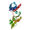
| ||||||||
|---|---|---|---|---|---|---|---|---|---|
| 1 | 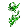
| ||||||||
| Unit cell |
|
-Components
| #1: Protein | Mass: 29655.859 Da / Num. of mol.: 1 Source method: isolated from a genetically manipulated source Source: (gene. exp.) Clostridium difficile (bacteria) / Gene: hadI / Production host: |
|---|---|
| #2: Chemical | ChemComp-SF4 /   Mass: 351.640 Da / Num. of mol.: 1 / Source method: obtained synthetically / Formula: Fe4S4 Mass: 351.640 Da / Num. of mol.: 1 / Source method: obtained synthetically / Formula: Fe4S4 |
-Experimental details
-Experiment
| Experiment | Method: X-RAY DIFFRACTION / Number of used crystals: 1 |
|---|
- Sample preparation
| Crystal | Density Matthews: 3.02 Å3/Da / Density % sol: 59.31 % |
|---|---|
| Crystal grow | Temperature: 289 K / Method: vapor diffusion, hanging drop / pH: 8.5 Details: 23 mg/mL protein in 50 mM MOPS, pH 7.2, 120 mM sodium chloride, 1.4 mM ADP, 9 mM magnesium chloride, 1 mM D-desthiobiotin, 6 mM DTT, 1.4 mM sodium dithionite, 100 mM sodium fluoride, 10 mM ...Details: 23 mg/mL protein in 50 mM MOPS, pH 7.2, 120 mM sodium chloride, 1.4 mM ADP, 9 mM magnesium chloride, 1 mM D-desthiobiotin, 6 mM DTT, 1.4 mM sodium dithionite, 100 mM sodium fluoride, 10 mM aluminum trifluoride, reservoir: 8% w/v PEG8000, 25% v/v MPD, 0.35 M sodium chloride, pH 8.5 (drop: 1 uL + 1 uL), VAPOR DIFFUSION, HANGING DROP, temperature 289K |
-Data collection
| Diffraction | Mean temperature: 100 K |
|---|---|
| Diffraction source | Source: SYNCHROTRON / Site: BESSY  / Beamline: 14.2 / Wavelength: 0.91841 Å / Beamline: 14.2 / Wavelength: 0.91841 Å |
| Detector | Type: RAYONIX MX-225 / Detector: CCD / Date: Jul 26, 2008 |
| Radiation | Monochromator: double crystal Si(111) / Protocol: SINGLE WAVELENGTH / Monochromatic (M) / Laue (L): M / Scattering type: x-ray |
| Radiation wavelength | Wavelength: 0.91841 Å / Relative weight: 1 |
| Reflection | Resolution: 3→30 Å / Num. all: 7197 / Num. obs: 7197 / % possible obs: 99.4 % / Observed criterion σ(F): 2 / Observed criterion σ(I): -3 / Redundancy: 7.3 % / Rsym value: 0.059 / Net I/σ(I): 21.32 |
| Reflection shell | Resolution: 3→3.1 Å / Redundancy: 7.6 % / Mean I/σ(I) obs: 4.35 / Rsym value: 0.486 / % possible all: 99.7 |
- Processing
| Software |
| |||||||||||||||||||||||||||||||||||||||||||||||||||||||||||||||||||||||||||
|---|---|---|---|---|---|---|---|---|---|---|---|---|---|---|---|---|---|---|---|---|---|---|---|---|---|---|---|---|---|---|---|---|---|---|---|---|---|---|---|---|---|---|---|---|---|---|---|---|---|---|---|---|---|---|---|---|---|---|---|---|---|---|---|---|---|---|---|---|---|---|---|---|---|---|---|---|
| Refinement | Method to determine structure: MOLECULAR REPLACEMENT Starting model: PDB ENTRY 4EHT Resolution: 3→19.672 Å / SU ML: 0.35 / σ(F): 1.99 / Phase error: 28.08 / Stereochemistry target values: ML
| |||||||||||||||||||||||||||||||||||||||||||||||||||||||||||||||||||||||||||
| Solvent computation | Shrinkage radii: 0.9 Å / VDW probe radii: 1.11 Å / Solvent model: FLAT BULK SOLVENT MODEL / Bsol: 49.682 Å2 / ksol: 0.265 e/Å3 | |||||||||||||||||||||||||||||||||||||||||||||||||||||||||||||||||||||||||||
| Displacement parameters |
| |||||||||||||||||||||||||||||||||||||||||||||||||||||||||||||||||||||||||||
| Refinement step | Cycle: LAST / Resolution: 3→19.672 Å
| |||||||||||||||||||||||||||||||||||||||||||||||||||||||||||||||||||||||||||
| Refine LS restraints |
| |||||||||||||||||||||||||||||||||||||||||||||||||||||||||||||||||||||||||||
| LS refinement shell |
| |||||||||||||||||||||||||||||||||||||||||||||||||||||||||||||||||||||||||||
| Refinement TLS params. | Method: refined / Refine-ID: X-RAY DIFFRACTION
| |||||||||||||||||||||||||||||||||||||||||||||||||||||||||||||||||||||||||||
| Refinement TLS group |
|
