- PDB-4ei0: Crystal structure of a DUF4466 family protein (PARMER_03218) from... -
+
Open data
ID or keywords:
Loading...
-
Basic information
Entry
Database: PDB / ID: 4ei0
Title
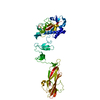
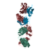
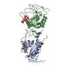
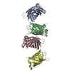
Crystal structure of a DUF4466 family protein (PARMER_03218) from Parabacteroides merdae ATCC 43184 at 2.00 A resolution
Components
uncharacterized hypothetical protein
Keywords
STRUCTURAL GENOMICS / UNKNOWN FUNCTION / PF14725 family protein / DUF4466 / Joint Center for Structural Genomics / JCSG / Protein Structure Initiative / PSI-BIOLOGY
Function / homology
Domain of unknown function DUF4466 / Domain of unknown function DUF4466 / PARMER_03128-like, N-terminal / Domain of unknown function (DUF4466) / Immunoglobulin-like / Sandwich / Mainly Beta / DUF4466 domain-containing protein
Function and homology information
Biological species
Parabacteroides merdae (bacteria)
Method
X-RAY DIFFRACTION / SYNCHROTRON / SAD / Resolution: 2 Å
Mass: 18.015 Da / Num. of mol.: 355 / Source method: isolated from a natural source / Formula: H2O
Has protein modification
Y
Sequence details
THIS CONSTRUCT (RESIDUES 23-333) WAS EXPRESSED WITH A PURIFICATION TAG MGSDKIHHHHHHENLYFQG. THE TAG ...THIS CONSTRUCT (RESIDUES 23-333) WAS EXPRESSED WITH A PURIFICATION TAG MGSDKIHHHHHHENLYFQG. THE TAG WAS REMOVED WITH TEV PROTEASE LEAVING ONLY A GLYCINE (0) FOLLOWED BY THE TARGET SEQUENCE.
-
Experimental details
-
Experiment
Experiment
Method: X-RAY DIFFRACTION / Number of used crystals: 1
-
Sample preparation
Crystal
Density Matthews: 2.5 Å3/Da / Density % sol: 50.74 %
Monochromator: double crystal Si(111) / Protocol: SINGLE WAVELENGTH / Monochromatic (M) / Laue (L): M / Scattering type: x-ray
Radiation wavelength
Wavelength: 0.9794 Å / Relative weight: 1
Reflection
Resolution: 2→29.469 Å / Num. obs: 47406 / % possible obs: 97.8 % / Observed criterion σ(I): -3 / Biso Wilson estimate: 32.338 Å2 / Rmerge(I) obs: 0.07 / Net I/σ(I): 9.67
Reflection shell
Diffraction-ID: 1
Resolution (Å)
Highest resolution (Å)
Rmerge(I) obs
Mean I/σ(I) obs
Num. measured obs
Num. unique obs
% possible all
2-2.07
0.706
1.6
22873
8642
97.5
2.07-2.15
0.515
2.2
23198
8706
98.3
2.15-2.25
0.406
2.8
23882
9123
98.3
2.25-2.37
0.303
3.5
22843
8991
97.5
2.37-2.52
0.235
4.6
24565
9084
98.8
2.52-2.71
0.167
6.1
22990
8750
98.4
2.71-2.99
0.1
9.6
23621
9125
97.8
2.99-3.42
0.059
15.7
23714
8852
98.3
3.42-4.3
0.036
23.2
22979
8785
97.1
4.3
0.029
27.4
23271
8838
96.5
-
Phasing
Phasing
Method: SAD
-
Processing
Software
Name
Version
Classification
NB
MolProbity
3beta29
modelbuilding
PDB_EXTRACT
3.1
dataextraction
SHELX
phasing
SHARP
phasing
XSCALE
December29, 2011
datascaling
REFMAC
5.6.0117
refinement
XDS
datareduction
SHELXD
phasing
Refinement
Method to determine structure: SAD / Resolution: 2→29.469 Å / Cor.coef. Fo:Fc: 0.967 / Cor.coef. Fo:Fc free: 0.953 / Occupancy max: 1 / Occupancy min: 0.5 / SU B: 7.887 / SU ML: 0.111 / Cross valid method: THROUGHOUT / σ(F): 0 / ESU R: 0.161 / ESU R Free: 0.148 Stereochemistry target values: MAXIMUM LIKELIHOOD WITH PHASES Details: 1.HYDROGENS HAVE BEEN ADDED IN THE RIDING POSITIONS. 2.A MET-INHIBITION PROTOCOL WAS USED FOR SELENOMETHIONINE INCORPORATION DURING PROTEIN EXPRESSION. THE OCCUPANCY OF THE SE ATOMS IN THE ...Details: 1.HYDROGENS HAVE BEEN ADDED IN THE RIDING POSITIONS. 2.A MET-INHIBITION PROTOCOL WAS USED FOR SELENOMETHIONINE INCORPORATION DURING PROTEIN EXPRESSION. THE OCCUPANCY OF THE SE ATOMS IN THE MSE RESIDUES WAS REDUCED TO 0.75 FOR THE REDUCED SCATTERING POWER DUE TO PARTIAL S-MET INCORPORATION. 3.ATOM RECORD CONTAINS SUM OF TLS AND RESIDUAL B FACTORS. ANISOU RECORD CONTAINS SUM OF TLS AND RESIDUAL U FACTORS. 4.WATERS WERE EXCLUDED FROM AUTOMATIC TLS ASSIGNMENT. 5.GLYCEROL (GOL) FROM THE CRYSTALLIZATION SOLUTION HAS BEEN MODELED IN THE SOLVENT STRUCTURE.
Rfactor
Num. reflection
% reflection
Selection details
Rfree
0.2161
2396
5.1 %
RANDOM
Rwork
0.1742
-
-
-
obs
0.1764
47345
99.12 %
-
Solvent computation
Ion probe radii: 0.8 Å / Shrinkage radii: 0.8 Å / VDW probe radii: 1.2 Å / Solvent model: BABINET MODEL WITH MASK
In the structure databanks used in Yorodumi, some data are registered as the other names, "COVID-19 virus" and "2019-nCoV". Here are the details of the virus and the list of structure data.
Jan 31, 2019. EMDB accession codes are about to change! (news from PDBe EMDB page)
EMDB accession codes are about to change! (news from PDBe EMDB page)
The allocation of 4 digits for EMDB accession codes will soon come to an end. Whilst these codes will remain in use, new EMDB accession codes will include an additional digit and will expand incrementally as the available range of codes is exhausted. The current 4-digit format prefixed with “EMD-” (i.e. EMD-XXXX) will advance to a 5-digit format (i.e. EMD-XXXXX), and so on. It is currently estimated that the 4-digit codes will be depleted around Spring 2019, at which point the 5-digit format will come into force.
The EM Navigator/Yorodumi systems omit the EMD- prefix.
Related info.:Q: What is EMD? / ID/Accession-code notation in Yorodumi/EM Navigator
Yorodumi is a browser for structure data from EMDB, PDB, SASBDB, etc.
This page is also the successor to EM Navigator detail page, and also detail information page/front-end page for Omokage search.
The word "yorodu" (or yorozu) is an old Japanese word meaning "ten thousand". "mi" (miru) is to see.
Related info.:EMDB / PDB / SASBDB / Comparison of 3 databanks / Yorodumi Search / Aug 31, 2016. New EM Navigator & Yorodumi / Yorodumi Papers / Jmol/JSmol / Function and homology information / Changes in new EM Navigator and Yorodumi
 Movie
Movie Controller

 Yorodumi
 Open data
 Basic information
 Components
 Keywords
 Function and homology information
 Parabacteroides merdae (bacteria)
 X-RAY DIFFRACTION /
  Authors
 Citation
 Structure visualization
 Downloads & links
 Other downloads
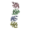


 PDBj
PDBj Assembly

 Mass: 92.094 Da / Num. of mol.: 1 / Source method: obtained synthetically / Formula: C3H8O3
 Mass: 18.015 Da / Num. of mol.: 355 / Source method: isolated from a natural source / Formula: H2O
 Sample preparation
 / Beamline: BL12-2 / Wavelength: 0.9794
  Processing