 Movie
Movie Controller

[English] 日本語
 Yorodumi
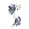
- PDB-4ehq: Crystal Structure of Calmodulin Binding Domain of Orai1 in Comple... -
+ Open data

- Basic information
| Entry | Database: PDB / ID: 4ehq | ||||||
|---|---|---|---|---|---|---|---|
| Title | Crystal Structure of Calmodulin Binding Domain of Orai1 in Complex with Ca2+/Calmodulin Displays a Unique Binding Mode | ||||||
 Components Components |
| ||||||
 Keywords Keywords | PROTEIN BINDING / calmodulin / Orai1 / calcium dependent inactivation / EF hand / Calcium binding / calcium-dependent inactivation / Calmodulin binding domain of Orai1 / none / cytosol | ||||||
| Function / homology |  Function and homology information Function and homology informationregulation of store-operated calcium channel activity / : / store-operated calcium entry / : / positive regulation of adenylate cyclase activity / regulation of response to tumor cell / positive regulation of autophagic cell death / DAPK1-calmodulin complex / store-operated calcium channel activity / : ...regulation of store-operated calcium channel activity / : / store-operated calcium entry / : / positive regulation of adenylate cyclase activity / regulation of response to tumor cell / positive regulation of autophagic cell death / DAPK1-calmodulin complex / store-operated calcium channel activity / : / : / : / : / : / mammary gland epithelium development / calcineurin-NFAT signaling cascade / calcium-ion regulated exocytosis / Elevation of cytosolic Ca2+ levels / transporter inhibitor activity / : / calcium ion import / type 3 metabotropic glutamate receptor binding / positive regulation of calcium ion transport / establishment of protein localization to membrane / positive regulation of DNA binding / negative regulation of high voltage-gated calcium channel activity / response to corticosterone / negative regulation of ryanodine-sensitive calcium-release channel activity / organelle localization by membrane tethering / ligand-gated ion channel signaling pathway / : / autophagosome membrane docking / regulation of synaptic vesicle exocytosis / negative regulation of calcium ion export across plasma membrane / regulation of ryanodine-sensitive calcium-release channel activity / regulation of cardiac muscle cell action potential / presynaptic endocytosis / regulation of calcium ion transport / calcineurin-mediated signaling / nitric-oxide synthase binding / regulation of cell communication by electrical coupling involved in cardiac conduction / adenylate cyclase binding / protein phosphatase activator activity / plasma membrane raft / regulation of synaptic vesicle endocytosis / detection of calcium ion / postsynaptic cytosol / regulation of cardiac muscle contraction / cell surface receptor signaling pathway via JAK-STAT / catalytic complex / positive regulation of nitric-oxide synthase activity / phosphatidylinositol 3-kinase binding / activation of adenylate cyclase activity / calcium channel inhibitor activity / presynaptic cytosol / enzyme regulator activity / cellular response to interferon-beta / regulation of release of sequestered calcium ion into cytosol by sarcoplasmic reticulum / Ion homeostasis / titin binding / regulation of cardiac muscle contraction by regulation of the release of sequestered calcium ion / regulation of calcium-mediated signaling / voltage-gated potassium channel complex / calcium channel complex / potassium ion transmembrane transport / regulation of heart rate / Antigen activates B Cell Receptor (BCR) leading to generation of second messengers / calyx of Held / nitric-oxide synthase regulator activity / adenylate cyclase activator activity / regulation of cytokinesis / protein serine/threonine kinase activator activity / cell periphery / spindle microtubule / sarcomere / positive regulation of receptor signaling pathway via JAK-STAT / response to amphetamine / calcium channel regulator activity / calcium-mediated signaling / response to calcium ion / cellular response to type II interferon / G2/M transition of mitotic cell cycle / Schaffer collateral - CA1 synapse / calcium ion transmembrane transport / calcium channel activity / spindle pole / disordered domain specific binding / positive regulation of insulin secretion / calcium-dependent protein binding / protein autophosphorylation / myelin sheath / synaptic vesicle membrane / growth cone / sperm midpiece / vesicle / basolateral plasma membrane / phospholipase C-activating G protein-coupled receptor signaling pathway / adaptive immune response / transmembrane transporter binding / calmodulin binding Similarity search - Function | ||||||
| Biological species |   Homo sapiens (human) Homo sapiens (human) | ||||||
| Method |  X-RAY DIFFRACTION / MOLECULAR REPLACEMENT / Resolution: 1.9005 Å X-RAY DIFFRACTION / MOLECULAR REPLACEMENT / Resolution: 1.9005 Å | ||||||
 Authors Authors | Liu, Y. / Zheng, X. / Mueller, G.A. / Sobhany, M. / DeRose, E.F. / Zhang, Y. / London, R.E. / Birnbaumer, L. | ||||||
 Citation Citation | Journal: J.Biol.Chem. / Year: 2012 Title: Crystal structure of calmodulin binding domain of orai1 in complex with ca2+*calmodulin displays a unique binding mode. Authors: Liu, Y. / Zheng, X. / Mueller, G.A. / Sobhany, M. / Derose, E.F. / Zhang, Y. / London, R.E. / Birnbaumer, L. | ||||||
| History |
|
- Structure visualization
| Structure viewer | Molecule: MolmilJmol/JSmol |
|---|
- Downloads & links
-Download
| PDBx/mmCIF format | 4ehq.cif.gz | 53 KB | Display | PDBx/mmCIF format |
|---|---|---|---|---|
| PDB format | pdb4ehq.ent.gz | 36.7 KB | Display | PDB format |
| PDBx/mmJSON format | 4ehq.json.gz | Tree view | PDBx/mmJSON format | |
| Others |  Other downloads Other downloads |
-Validation report
| Arichive directory | https://data.pdbj.org/pub/pdb/validation_reports/eh/4ehqftp://data.pdbj.org/pub/pdb/validation_reports/eh/4ehq | HTTPS FTP |
|---|
-Related structure data
| Related structure data | 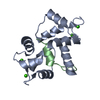 1iwqS S: Starting model for refinement |
|---|---|
| Similar structure data |


-Links
 PDBj
PDBj


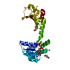
- Assembly
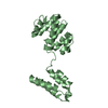
| Deposited unit | 
| ||||||||
|---|---|---|---|---|---|---|---|---|---|
| 1 |
| ||||||||
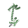
| Unit cell |
|
-Components
| #1: Protein | Mass: 16721.350 Da / Num. of mol.: 1 Source method: isolated from a genetically manipulated source Source: (gene. exp.) Gene: Calm1, Calm, Cam, Cam1, Calm2, Cam2, Camb, Calm3, Cam3, Camc Production host:  | ||||
|---|---|---|---|---|---|
| #2: Protein/peptide | Mass: 2393.895 Da / Num. of mol.: 1 / Source method: obtained synthetically / Details: Synthetic peptide / Source: (synth.) Homo sapiens (human) / References: UniProt: Q96D31 | ||||
| #3: Chemical | ChemComp-CA /   Mass: 40.078 Da / Num. of mol.: 4 / Source method: obtained synthetically / Formula: Ca Mass: 40.078 Da / Num. of mol.: 4 / Source method: obtained synthetically / Formula: Ca#4: Chemical | ChemComp-GBL / 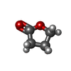  Mass: 86.089 Da / Num. of mol.: 4 / Source method: obtained synthetically / Formula: C4H6O2 Mass: 86.089 Da / Num. of mol.: 4 / Source method: obtained synthetically / Formula: C4H6O2#5: Water | ChemComp-HOH / |  Mass: 18.015 Da / Num. of mol.: 88 / Source method: isolated from a natural source / Formula: H2O Mass: 18.015 Da / Num. of mol.: 88 / Source method: isolated from a natural source / Formula: H2O |
-Experimental details
-Experiment
| Experiment | Method: X-RAY DIFFRACTION / Number of used crystals: 1 |
|---|
- Sample preparation
| Crystal | Density Matthews: 1.94 Å3/Da / Density % sol: 36.57 % |
|---|---|
| Crystal grow | Temperature: 293 K / Method: vapor diffusion, hanging drop / pH: 6 Details: 0.1 M Bis-Tris pH 6.0, 40% PPG P400, 14% butyrolactone, 1% n-octyl-beta-D-glucopyranoside, VAPOR DIFFUSION, HANGING DROP, temperature 293K |
-Data collection
| Diffraction | Mean temperature: 100 K |
|---|---|
| Diffraction source | Source: ROTATING ANODE / Type: RIGAKU MICROMAX-007 HF / Wavelength: 1.5418 Å |
| Detector | Type: RIGAKU SATURN 92 / Detector: CCD / Date: Aug 23, 2010 |
| Radiation | Monochromator: copper anode / Protocol: SINGLE WAVELENGTH / Monochromatic (M) / Laue (L): M / Scattering type: x-ray |
| Radiation wavelength | Wavelength: 1.5418 Å / Relative weight: 1 |
| Reflection | Resolution: 1.9→50 Å / Num. all: 11939 / Num. obs: 11497 / % possible obs: 96.3 % / Observed criterion σ(F): 1 / Observed criterion σ(I): 1 / Redundancy: 3.4 % / Biso Wilson estimate: 18 Å2 / Rsym value: 0.084 / Net I/σ(I): 12.9 |
| Reflection shell | Resolution: 1.9→1.97 Å / Redundancy: 2.2 % / Rmerge(I) obs: 0.231 / Mean I/σ(I) obs: 3.9 / Num. unique all: 853 / % possible all: 73.9 |
- Processing
| Software |
| |||||||||||||||||||||||||||||||||||
|---|---|---|---|---|---|---|---|---|---|---|---|---|---|---|---|---|---|---|---|---|---|---|---|---|---|---|---|---|---|---|---|---|---|---|---|---|
| Refinement | Method to determine structure: MOLECULAR REPLACEMENT Starting model: PDB ENTRY 1IWQ Resolution: 1.9005→19.987 Å / SU ML: 0.21 / σ(F): 0 / σ(I): 0 / Phase error: 19.33 / Stereochemistry target values: MLHL
| |||||||||||||||||||||||||||||||||||
| Solvent computation | Shrinkage radii: 0.9 Å / VDW probe radii: 1.11 Å / Solvent model: FLAT BULK SOLVENT MODEL / Bsol: 44.125 Å2 / ksol: 0.415 e/Å3 | |||||||||||||||||||||||||||||||||||
| Displacement parameters |
| |||||||||||||||||||||||||||||||||||
| Refinement step | Cycle: LAST / Resolution: 1.9005→19.987 Å
| |||||||||||||||||||||||||||||||||||
| Refine LS restraints |
| |||||||||||||||||||||||||||||||||||
| LS refinement shell |
|
