 Movie
Movie Controller

[English] 日本語
 Yorodumi
- PDB-4edt: The structure of the S. aureus DnaG RNA Polymerase Domain bound t... -
+ Open data

- Basic information
| Entry | Database: PDB / ID: 4edt | ||||||
|---|---|---|---|---|---|---|---|
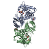
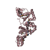
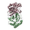
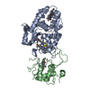
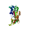
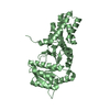
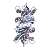
| Title | The structure of the S. aureus DnaG RNA Polymerase Domain bound to ppGpp and Manganese | ||||||
 Components Components | DNA primase | ||||||
 Keywords Keywords | transferase/transferase inhibitor / Catalytic Domain / Bacterial / nucleotide / nucleoside triphosphate / nucleoside polyphosphate / protein-ligand complex / transferase / transferase-transferase inhibitor complex | ||||||
| Function / homology |  Function and homology information Function and homology informationDNA primase DnaG / primosome complex / DNA replication, synthesis of primer / DNA helicase activity / DNA-directed RNA polymerase complex / DNA-directed RNA polymerase activity / DNA binding / zinc ion binding / ATP binding / cytoplasm Similarity search - Function | ||||||
| Biological species |   Staphylococcus aureus (bacteria) Staphylococcus aureus (bacteria) | ||||||
| Method |  X-RAY DIFFRACTION / SYNCHROTRON / MOLECULAR REPLACEMENT / Resolution: 2.005 Å X-RAY DIFFRACTION / SYNCHROTRON / MOLECULAR REPLACEMENT / Resolution: 2.005 Å | ||||||
 Authors Authors | Rymer, R.U. / Solorio, F.A. / Chu, C. / Corn, J.E. / Wang, J.D. / Berger, J.M. | ||||||
 Citation Citation | Journal: Structure / Year: 2012 Title: Binding Mechanism of Metal-NTP Substrates and Stringent-Response Alarmones to Bacterial DnaG-Type Primases. Authors: Rymer, R.U. / Solorio, F.A. / Tehranchi, A.K. / Chu, C. / Corn, J.E. / Keck, J.L. / Wang, J.D. / Berger, J.M. | ||||||
| History |
|
- Structure visualization
| Structure viewer | Molecule: MolmilJmol/JSmol |
|---|
- Downloads & links
-Download
| PDBx/mmCIF format | 4edt.cif.gz | 154.3 KB | Display | PDBx/mmCIF format |
|---|---|---|---|---|
| PDB format | pdb4edt.ent.gz | 120.5 KB | Display | PDB format |
| PDBx/mmJSON format | 4edt.json.gz | Tree view | PDBx/mmJSON format | |
| Others |  Other downloads Other downloads |
-Validation report
| Arichive directory | https://data.pdbj.org/pub/pdb/validation_reports/ed/4edtftp://data.pdbj.org/pub/pdb/validation_reports/ed/4edt | HTTPS FTP |
|---|
-Related structure data
| Related structure data | 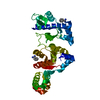 4e2kC  4edgC 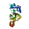 4edkC 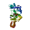 4edrC  4edvC 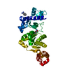 4ee1C C: citing same article ( |
|---|---|
| Similar structure data |


-Links
 PDBj
PDBj


- Assembly
| Deposited unit | 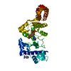
| ||||||||
|---|---|---|---|---|---|---|---|---|---|
| 1 |
| ||||||||
| Unit cell |
| ||||||||
| Details | monomer |
-Components
| #1: Protein | Mass: 37677.508 Da / Num. of mol.: 1 / Fragment: unp residues 111-436 Source method: isolated from a genetically manipulated source Source: (gene. exp.) Staphylococcus aureus (bacteria) / Gene: dnaG / Plasmid: pET28b / Production host: References: UniProt: O05338, Transferases; Transferring phosphorus-containing groups; Nucleotidyltransferases | ||
|---|---|---|---|
| #2: Chemical | ChemComp-BEN /   Mass: 120.152 Da / Num. of mol.: 1 / Source method: obtained synthetically / Formula: C7H8N2 Mass: 120.152 Da / Num. of mol.: 1 / Source method: obtained synthetically / Formula: C7H8N2 | ||
| #3: Chemical | ChemComp-G4P /   Type: RNA linking / Mass: 603.160 Da / Num. of mol.: 1 / Source method: obtained synthetically / Formula: C10H17N5O17P4 Type: RNA linking / Mass: 603.160 Da / Num. of mol.: 1 / Source method: obtained synthetically / Formula: C10H17N5O17P4 | ||
| #4: Chemical |   Mass: 54.938 Da / Num. of mol.: 2 / Source method: obtained synthetically / Formula: Mn Mass: 54.938 Da / Num. of mol.: 2 / Source method: obtained synthetically / Formula: Mn#5: Water | ChemComp-HOH / |  Mass: 18.015 Da / Num. of mol.: 338 / Source method: isolated from a natural source / Formula: H2O Mass: 18.015 Da / Num. of mol.: 338 / Source method: isolated from a natural source / Formula: H2O |
-Experimental details
-Experiment
| Experiment | Method: X-RAY DIFFRACTION / Number of used crystals: 1 |
|---|
- Sample preparation
| Crystal | Density Matthews: 3.43 Å3/Da / Density % sol: 64.12 % |
|---|---|
| Crystal grow | Temperature: 292 K / Method: vapor diffusion, hanging drop / pH: 6.5 Details: 0.15M sodium thiocyanate, 0.1M Bis-Tris, 13% PEG3350, 2% Benzamidine, 2.5 mM ppGpp, 10 mM MnCl2, 1 mM MgCl2, pH 6.5, VAPOR DIFFUSION, HANGING DROP, temperature 292K |
-Data collection
| Diffraction | Mean temperature: 100 K |
|---|---|
| Diffraction source | Source: SYNCHROTRON / Site: ALS  / Beamline: 8.3.1 / Wavelength: 1.115 Å / Beamline: 8.3.1 / Wavelength: 1.115 Å |
| Detector | Type: ADSC QUANTUM 315r / Detector: CCD / Date: Nov 20, 2009 |
| Radiation | Monochromator: Double flat crystal, Si(111) / Protocol: SINGLE WAVELENGTH / Monochromatic (M) / Laue (L): M / Scattering type: x-ray |
| Radiation wavelength | Wavelength: 1.115 Å / Relative weight: 1 |
| Reflection | Resolution: 2.005→131.405 Å / Num. all: 34635 / Num. obs: 34635 / % possible obs: 99.7 % / Redundancy: 8.2 % / Rsym value: 0.075 |
| Reflection shell | Resolution: 2→2.11 Å / Redundancy: 7.8 % / Mean I/σ(I) obs: 3.2 / Rsym value: 0.63 / % possible all: 98.1 |
- Processing
| Software |
| |||||||||||||||||||||||||||||||||||||||||||||||||||||||||||||||||||||||||||||||||||||||||||||||||||||||||||||||||||||||||||||
|---|---|---|---|---|---|---|---|---|---|---|---|---|---|---|---|---|---|---|---|---|---|---|---|---|---|---|---|---|---|---|---|---|---|---|---|---|---|---|---|---|---|---|---|---|---|---|---|---|---|---|---|---|---|---|---|---|---|---|---|---|---|---|---|---|---|---|---|---|---|---|---|---|---|---|---|---|---|---|---|---|---|---|---|---|---|---|---|---|---|---|---|---|---|---|---|---|---|---|---|---|---|---|---|---|---|---|---|---|---|---|---|---|---|---|---|---|---|---|---|---|---|---|---|---|---|---|
| Refinement | Method to determine structure: MOLECULAR REPLACEMENT / Resolution: 2.005→43.926 Å / Occupancy max: 1 / Occupancy min: 0.29 / SU ML: 0.22 / σ(F): 1.35 / Phase error: 20.72 / Stereochemistry target values: ML
| |||||||||||||||||||||||||||||||||||||||||||||||||||||||||||||||||||||||||||||||||||||||||||||||||||||||||||||||||||||||||||||
| Solvent computation | Shrinkage radii: 0.98 Å / VDW probe radii: 1.2 Å / Solvent model: FLAT BULK SOLVENT MODEL / Bsol: 36.086 Å2 / ksol: 0.322 e/Å3 | |||||||||||||||||||||||||||||||||||||||||||||||||||||||||||||||||||||||||||||||||||||||||||||||||||||||||||||||||||||||||||||
| Displacement parameters | Biso max: 148.06 Å2 / Biso mean: 36.3554 Å2 / Biso min: 13.27 Å2
| |||||||||||||||||||||||||||||||||||||||||||||||||||||||||||||||||||||||||||||||||||||||||||||||||||||||||||||||||||||||||||||
| Refinement step | Cycle: LAST / Resolution: 2.005→43.926 Å
| |||||||||||||||||||||||||||||||||||||||||||||||||||||||||||||||||||||||||||||||||||||||||||||||||||||||||||||||||||||||||||||
| Refine LS restraints |
| |||||||||||||||||||||||||||||||||||||||||||||||||||||||||||||||||||||||||||||||||||||||||||||||||||||||||||||||||||||||||||||
| LS refinement shell | Refine-ID: X-RAY DIFFRACTION / Total num. of bins used: 13
| |||||||||||||||||||||||||||||||||||||||||||||||||||||||||||||||||||||||||||||||||||||||||||||||||||||||||||||||||||||||||||||
| Refinement TLS params. | Method: refined / Refine-ID: X-RAY DIFFRACTION
| |||||||||||||||||||||||||||||||||||||||||||||||||||||||||||||||||||||||||||||||||||||||||||||||||||||||||||||||||||||||||||||
| Refinement TLS group |
|
