 Movie
Movie Controller

[English] 日本語
 Yorodumi
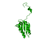
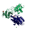
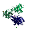
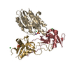
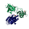
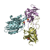
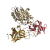
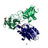
- PDB-4ec2: Crystal structure of trimeric frataxin from the yeast Saccharomyc... -
+ Open data

- Basic information
| Entry | Database: PDB / ID: 4ec2 | ||||||
|---|---|---|---|---|---|---|---|
| Title | Crystal structure of trimeric frataxin from the yeast Saccharomyces cerevisiae, complexed with ferrous | ||||||
 Components Components | Frataxin homolog, mitochondrial | ||||||
 Keywords Keywords | TRANSPORT PROTEIN / alpha/beta sandwich / metallochaperone / iron-storage | ||||||
| Function / homology |  Function and homology information Function and homology informationMitochondrial iron-sulfur cluster biogenesis / Mitochondrial protein import / Maturation of TCA enzymes and regulation of TCA cycle / Complex III assembly / mitochondrial electron transport, succinate to ubiquinone / iron chaperone activity / iron-sulfur cluster assembly complex / heme biosynthetic process / iron-sulfur cluster assembly / response to iron(II) ion ...Mitochondrial iron-sulfur cluster biogenesis / Mitochondrial protein import / Maturation of TCA enzymes and regulation of TCA cycle / Complex III assembly / mitochondrial electron transport, succinate to ubiquinone / iron chaperone activity / iron-sulfur cluster assembly complex / heme biosynthetic process / iron-sulfur cluster assembly / response to iron(II) ion / ferroxidase / ferroxidase activity / ferric iron binding / glutathione metabolic process / iron ion transport / ferrous iron binding / mitochondrial intermembrane space / 2 iron, 2 sulfur cluster binding / response to oxidative stress / intracellular iron ion homeostasis / mitochondrial inner membrane / mitochondrial matrix / mitochondrion / identical protein binding Similarity search - Function | ||||||
| Biological species |  | ||||||
| Method |  X-RAY DIFFRACTION / SYNCHROTRON / MOLECULAR REPLACEMENT / Resolution: 3.002 Å X-RAY DIFFRACTION / SYNCHROTRON / MOLECULAR REPLACEMENT / Resolution: 3.002 Å | ||||||
 Authors Authors | Soderberg, C.A.G. / Rajan, S. / Gakh, O. / Isaya, G. / Al-Karadaghi, S. | ||||||
 Citation Citation | Journal: J.Biol.Chem. / Year: 2013 Title: The molecular basis of iron-induced oligomerization of frataxin and the role of the ferroxidation reaction in oligomerization. Authors: Soderberg, C.A. / Rajan, S. / Shkumatov, A.V. / Gakh, O. / Schaefer, S. / Ahlgren, E.C. / Svergun, D.I. / Isaya, G. / Al-Karadaghi, S. #1: Journal: J.Mol.Biol. / Year: 2011Title: Oligomerization propensity and flexibility of yeast frataxin studied by x-ray crystallography and small-angle x-ray scattering Authors: Soderberg, C.A.G. / Shkumatov, A.V. / Rajan, S. / Gakh, O. / Svergun, D.I. / Isaya, G. / Al-karadaghi, S. #2: Journal: Structure / Year: 2006Title: The structures of frataxin oligomers reveal the mechanism for the delivery and detoxification of iron Authors: Karlberg, T. / Schagerlof, U. / Gakh, O. / Park, S. / Ryde, U. / Lindahl, M. / Leath, K. / Garman, E. / Isaya, G. / Al-karadaghi, S. | ||||||
| History |
| ||||||
| Remark 650 | HELIX DETERMINATION METHOD: AUTHOR | ||||||
| Remark 700 | SHEET DETERMINATION METHOD: AUTHOR |
- Structure visualization
| Structure viewer | Molecule: MolmilJmol/JSmol |
|---|
- Downloads & links
-Download
| PDBx/mmCIF format | 4ec2.cif.gz | 36.1 KB | Display | PDBx/mmCIF format |
|---|---|---|---|---|
| PDB format | pdb4ec2.ent.gz | 24.4 KB | Display | PDB format |
| PDBx/mmJSON format | 4ec2.json.gz | Tree view | PDBx/mmJSON format | |
| Others |  Other downloads Other downloads |
-Validation report
| Arichive directory | https://data.pdbj.org/pub/pdb/validation_reports/ec/4ec2ftp://data.pdbj.org/pub/pdb/validation_reports/ec/4ec2 | HTTPS FTP |
|---|
-Related structure data
| Related structure data |  2fqlS S: Starting model for refinement |
|---|---|
| Similar structure data |


-Links
 PDBj
PDBj


- Assembly
| Deposited unit | 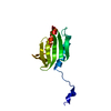
| ||||||||
|---|---|---|---|---|---|---|---|---|---|
| 1 | 
| ||||||||
| Unit cell |
|
-Components
| #1: Protein | Mass: 13671.182 Da / Num. of mol.: 1 / Fragment: UNP residues 52-174 / Mutation: Y73A Source method: isolated from a genetically manipulated source Source: (gene. exp.) Strain: ATCC 204508 / S288c / Gene: YFH1, YDL120W / Production host:  |
|---|---|
| #2: Chemical | ChemComp-FE2 /   Mass: 55.845 Da / Num. of mol.: 1 / Source method: obtained synthetically / Formula: Fe Mass: 55.845 Da / Num. of mol.: 1 / Source method: obtained synthetically / Formula: Fe |
-Experimental details
-Experiment
| Experiment | Method: X-RAY DIFFRACTION / Number of used crystals: 1 |
|---|
- Sample preparation
| Crystal | Density Matthews: 5.48 Å3/Da / Density % sol: 77.54 % |
|---|---|
| Crystal grow | Temperature: 288 K / Method: vapor diffusion, hanging drop / pH: 8.5 Details: 1.7 M (NH4)2SO4, 0.2 M Li2SO4, 4 % Gamma-butyrolactone, 0.1 M Tris pH 8.5, VAPOR DIFFUSION, HANGING DROP, temperature 288K |
-Data collection
| Diffraction | Mean temperature: 100 K |
|---|---|
| Diffraction source | Source: SYNCHROTRON / Site: MAX II  / Beamline: I911-2 / Wavelength: 1.038 Å / Beamline: I911-2 / Wavelength: 1.038 Å |
| Detector | Type: MAR CCD 165 mm / Detector: CCD / Date: Sep 10, 2008 |
| Radiation | Monochromator: SI (111) DOUBLE CRYSTAL / Protocol: SINGLE WAVELENGTH / Monochromatic (M) / Laue (L): M / Scattering type: x-ray |
| Radiation wavelength | Wavelength: 1.038 Å / Relative weight: 1 |
| Reflection | Resolution: 3→50 Å / Num. obs: 11254 / % possible obs: 96.9 % / Redundancy: 3.32 % / Rmerge(I) obs: 0.064 / Net I/σ(I): 11.62 |
| Reflection shell | Resolution: 3→3.08 Å / Redundancy: 3.12 % / Rmerge(I) obs: 0.632 / Mean I/σ(I) obs: 1.76 / Num. unique all: 791 / % possible all: 93.3 |
- Processing
| Software |
| |||||||||||||||||||||||||||||||||||||||||||||||||||||||||||||||
|---|---|---|---|---|---|---|---|---|---|---|---|---|---|---|---|---|---|---|---|---|---|---|---|---|---|---|---|---|---|---|---|---|---|---|---|---|---|---|---|---|---|---|---|---|---|---|---|---|---|---|---|---|---|---|---|---|---|---|---|---|---|---|---|---|
| Refinement | Method to determine structure: MOLECULAR REPLACEMENT Starting model: 2FQL Resolution: 3.002→24.815 Å / SU ML: 1.16 / σ(F): 1.29 / Phase error: 39.05 / Stereochemistry target values: ML
| |||||||||||||||||||||||||||||||||||||||||||||||||||||||||||||||
| Solvent computation | Shrinkage radii: 0.86 Å / VDW probe radii: 1.1 Å / Solvent model: FLAT BULK SOLVENT MODEL / Bsol: 59.031 Å2 / ksol: 0.287 e/Å3 | |||||||||||||||||||||||||||||||||||||||||||||||||||||||||||||||
| Refinement step | Cycle: LAST / Resolution: 3.002→24.815 Å
| |||||||||||||||||||||||||||||||||||||||||||||||||||||||||||||||
| Refine LS restraints |
| |||||||||||||||||||||||||||||||||||||||||||||||||||||||||||||||
| LS refinement shell |
|
