 Movie
Movie Controller

[English] 日本語
 Yorodumi
- PDB-4ea5: Structure of the glycoslyase domain of MBD4 bound to a 5hmU conta... -
+ Open data

- Basic information
| Entry | Database: PDB / ID: 4ea5 | ||||||
|---|---|---|---|---|---|---|---|
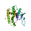
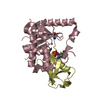
| Title | Structure of the glycoslyase domain of MBD4 bound to a 5hmU containing DNA | ||||||
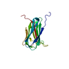
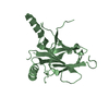
 Components Components |
| ||||||
 Keywords Keywords | HYDROLASE/DNA / HhH DNA glycosylase family / HYDROLASE-DNA complex | ||||||
| Function / homology |  Function and homology information Function and homology informationsatellite DNA binding / pyrimidine-specific mismatch base pair DNA N-glycosylase activity / depyrimidination / DNA N-glycosylase activity / Displacement of DNA glycosylase by APEX1 / Hydrolases; Glycosylases; Hydrolysing N-glycosyl compounds / Recognition and association of DNA glycosylase with site containing an affected pyrimidine / Cleavage of the damaged pyrimidine / DNA endonuclease activity / response to estradiol ...satellite DNA binding / pyrimidine-specific mismatch base pair DNA N-glycosylase activity / depyrimidination / DNA N-glycosylase activity / Displacement of DNA glycosylase by APEX1 / Hydrolases; Glycosylases; Hydrolysing N-glycosyl compounds / Recognition and association of DNA glycosylase with site containing an affected pyrimidine / Cleavage of the damaged pyrimidine / DNA endonuclease activity / response to estradiol / nuclear speck / DNA repair / DNA binding / nucleoplasm / nucleus Similarity search - Function | ||||||
| Biological species |  Homo sapiens (human) Homo sapiens (human) | ||||||
| Method |  X-RAY DIFFRACTION / SYNCHROTRON / MOLECULAR REPLACEMENT / Resolution: 2.14 Å X-RAY DIFFRACTION / SYNCHROTRON / MOLECULAR REPLACEMENT / Resolution: 2.14 Å | ||||||
 Authors Authors | Morera, S. / Vigouroux, A. | ||||||
 Citation Citation | Journal: Nucleic Acids Res. / Year: 2012 Title: Biochemical and structural characterization of the glycosylase domain of MBD4 bound to thymine and 5-hydroxymethyuracil-containing DNA. Authors: Morera, S. / Grin, I. / Vigouroux, A. / Couve, S. / Henriot, V. / Saparbaev, M. / Ishchenko, A.A. | ||||||
| History |
|
- Structure visualization
| Structure viewer | Molecule: MolmilJmol/JSmol |
|---|
- Downloads & links
-Download
| PDBx/mmCIF format | 4ea5.cif.gz | 58.4 KB | Display | PDBx/mmCIF format |
|---|---|---|---|---|
| PDB format | pdb4ea5.ent.gz | 38.2 KB | Display | PDB format |
| PDBx/mmJSON format | 4ea5.json.gz | Tree view | PDBx/mmJSON format | |
| Others |  Other downloads Other downloads |
-Validation report
| Arichive directory | https://data.pdbj.org/pub/pdb/validation_reports/ea/4ea5ftp://data.pdbj.org/pub/pdb/validation_reports/ea/4ea5 | HTTPS FTP |
|---|
-Related structure data
| Related structure data |  4e9eSC  4e9fC 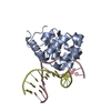 4e9gC  4e9hC 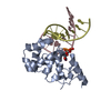 4ea4C S: Starting model for refinement C: citing same article ( |
|---|---|
| Similar structure data |


-Links
 PDBj
PDBj


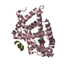
- Assembly
| Deposited unit | 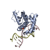
| ||||||||
|---|---|---|---|---|---|---|---|---|---|
| 1 |
| ||||||||
| Unit cell |
|
-Components
| #1: Protein | Mass: 19234.236 Da / Num. of mol.: 1 / Fragment: glycosylase domain (residues 426-580) of MBD4 / Mutation: D560A Source method: isolated from a genetically manipulated source Source: (gene. exp.) Homo sapiens (human) / Gene: MBD4, MED1 / Production host:  References: UniProt: O95243, Hydrolases; Glycosylases; Hydrolysing N-glycosyl compounds |
|---|---|
| #2: DNA chain | Mass: 3664.381 Da / Num. of mol.: 1 / Source method: obtained synthetically / Details: Synthetic DNA |
| #3: DNA chain | Mass: 3695.390 Da / Num. of mol.: 1 / Source method: obtained synthetically / Details: Synthetic DNA |
| #4: Water | ChemComp-HOH /  Mass: 18.015 Da / Num. of mol.: 71 / Source method: isolated from a natural source / Formula: H2O Mass: 18.015 Da / Num. of mol.: 71 / Source method: isolated from a natural source / Formula: H2O |
-Experimental details
-Experiment
| Experiment | Method: X-RAY DIFFRACTION / Number of used crystals: 1 |
|---|
- Sample preparation
| Crystal | Density Matthews: 1.98 Å3/Da / Density % sol: 37.94 % |
|---|---|
| Crystal grow | Temperature: 298 K / Method: vapor diffusion, hanging drop / pH: 8.5 Details: 20% Jeffamine M2070, 20% dimethylsulfoxite, pH 8.5, VAPOR DIFFUSION, HANGING DROP, temperature 298K |
-Data collection
| Diffraction | Mean temperature: 100 K |
|---|---|
| Diffraction source | Source: SYNCHROTRON / Site: SOLEIL  / Beamline: PROXIMA 1 / Wavelength: 0.9 Å / Beamline: PROXIMA 1 / Wavelength: 0.9 Å |
| Detector | Type: PSI PILATUS 6M / Detector: PIXEL / Date: Nov 27, 2011 |
| Radiation | Monochromator: Si 111 / Protocol: SINGLE WAVELENGTH / Monochromatic (M) / Laue (L): M / Scattering type: x-ray |
| Radiation wavelength | Wavelength: 0.9 Å / Relative weight: 1 |
| Reflection | Resolution: 2.14→47.13 Å / Num. all: 12000 / Num. obs: 11828 / % possible obs: 99.2 % / Observed criterion σ(F): 2 / Observed criterion σ(I): 2 |
| Reflection shell | Resolution: 2.14→2.27 Å / % possible all: 95.6 |
- Processing
| Software |
| ||||||||||||||||||||||||||||||||||||||||||||||||||||||||||||||||||||||||||||||||||||||||||||||||||||||||||||||||||
|---|---|---|---|---|---|---|---|---|---|---|---|---|---|---|---|---|---|---|---|---|---|---|---|---|---|---|---|---|---|---|---|---|---|---|---|---|---|---|---|---|---|---|---|---|---|---|---|---|---|---|---|---|---|---|---|---|---|---|---|---|---|---|---|---|---|---|---|---|---|---|---|---|---|---|---|---|---|---|---|---|---|---|---|---|---|---|---|---|---|---|---|---|---|---|---|---|---|---|---|---|---|---|---|---|---|---|---|---|---|---|---|---|---|---|---|
| Refinement | Method to determine structure: MOLECULAR REPLACEMENT Starting model: PDB ENTRY 4E9E Resolution: 2.14→47.13 Å / Cor.coef. Fo:Fc: 0.9264 / Cor.coef. Fo:Fc free: 0.9118 / SU R Cruickshank DPI: 0.227 / Cross valid method: THROUGHOUT / σ(F): 0 / Stereochemistry target values: Engh & Huber
| ||||||||||||||||||||||||||||||||||||||||||||||||||||||||||||||||||||||||||||||||||||||||||||||||||||||||||||||||||
| Refine analyze | Luzzati coordinate error obs: 0.226 Å | ||||||||||||||||||||||||||||||||||||||||||||||||||||||||||||||||||||||||||||||||||||||||||||||||||||||||||||||||||
| Refinement step | Cycle: LAST / Resolution: 2.14→47.13 Å
| ||||||||||||||||||||||||||||||||||||||||||||||||||||||||||||||||||||||||||||||||||||||||||||||||||||||||||||||||||
| Refine LS restraints |
| ||||||||||||||||||||||||||||||||||||||||||||||||||||||||||||||||||||||||||||||||||||||||||||||||||||||||||||||||||
| LS refinement shell | Resolution: 2.14→2.34 Å / Total num. of bins used: 6
|
