[English] 日本語
Yorodumi
- PDB-4e93: Crystal structure of human Feline Sarcoma Viral Oncogene Homologu... -

+
Open data


ID or keywords:

Loading...

-
Basic information

Entry
Database: PDB / ID: 4.0E+93
TitleCrystal structure of human Feline Sarcoma Viral Oncogene Homologue (v-FES)in complex with TAE684
ComponentsTyrosine-protein kinase Fes/Fps
KeywordsTRANSFERASE/TRANSFERASE INHIBITOR / V-FES / FUJINAMI / AVIAN SARCOMA / VIRAL / ONCOGENE / FELINE SARCOMA VIRUS / ATP-BINDING / KINASE / NUCLEOTIDE-BINDING / PHOSPHOPROTEIN / PROTO-ONCOGENE / SH2 DOMAIN / TRANSFERASE / TYROSINE-PROTEIN KINASE / TRANSFERASE-TRANSFERASE INHIBITOR complex / Structural Genomics / Structural Genomics Consortium / SGC
Function / homology
Function and homology information


positive regulation of myeloid cell differentiation / regulation of mast cell degranulation / regulation of vesicle-mediated transport / cellular response to vitamin D / SEMA3A-Plexin repulsion signaling by inhibiting Integrin adhesion / CRMPs in Sema3A signaling / microtubule bundle formation / positive regulation of monocyte differentiation / regulation of cell motility / centrosome cycle ...positive regulation of myeloid cell differentiation / regulation of mast cell degranulation / regulation of vesicle-mediated transport / cellular response to vitamin D / SEMA3A-Plexin repulsion signaling by inhibiting Integrin adhesion / CRMPs in Sema3A signaling / microtubule bundle formation / positive regulation of monocyte differentiation / regulation of cell motility / centrosome cycle / myoblast proliferation / cardiac muscle cell proliferation / regulation of cell differentiation / Sema3A PAK dependent Axon repulsion / regulation of cell adhesion / immunoglobulin receptor binding / positive regulation of microtubule polymerization / phosphatidylinositol binding / non-specific protein-tyrosine kinase / non-membrane spanning protein tyrosine kinase activity / peptidyl-tyrosine phosphorylation / Signaling by SCF-KIT / cytoplasmic side of plasma membrane / positive regulation of neuron projection development / chemotaxis / microtubule cytoskeleton / regulation of cell population proliferation / regulation of cell shape / protein tyrosine kinase activity / cytoplasmic vesicle / microtubule binding / protein autophosphorylation / cell adhesion / focal adhesion / Golgi apparatus / ATP binding / plasma membrane / cytoplasm / cytosol
Similarity search - Function
Tyrosine-protein kinase, Fes/Fps type / Fes/Fps/Fer, SH2 domain / Fes/CIP4, and EFC/F-BAR homology domain / Fes/CIP4 homology domain / FCH domain / F-BAR domain / F-BAR domain profile. / AH/BAR domain superfamily / SH2 domain / SHC Adaptor Protein ...Tyrosine-protein kinase, Fes/Fps type / Fes/Fps/Fer, SH2 domain / Fes/CIP4, and EFC/F-BAR homology domain / Fes/CIP4 homology domain / FCH domain / F-BAR domain / F-BAR domain profile. / AH/BAR domain superfamily / SH2 domain / SHC Adaptor Protein / : / SH2 domain / Src homology 2 (SH2) domain profile. / Src homology 2 domains / SH2 domain / SH2 domain superfamily / Tyrosine-protein kinase, catalytic domain / Tyrosine kinase, catalytic domain / Tyrosine protein kinases specific active-site signature. / Tyrosine-protein kinase, active site / Serine-threonine/tyrosine-protein kinase, catalytic domain / Protein tyrosine and serine/threonine kinase / Transferase(Phosphotransferase) domain 1 / Transferase(Phosphotransferase); domain 1 / Phosphorylase Kinase; domain 1 / Phosphorylase Kinase; domain 1 / Protein kinase, ATP binding site / Protein kinases ATP-binding region signature. / Protein kinase domain profile. / Protein kinase domain / Protein kinase-like domain superfamily / 2-Layer Sandwich / Orthogonal Bundle / Mainly Alpha / Alpha Beta
Similarity search - Domain/homology
Chem-GUI / Tyrosine-protein kinase Fes/Fps
Similarity search - Component
Biological speciesHomo sapiens (human)
MethodX-RAY DIFFRACTION / MOLECULAR REPLACEMENT / molecular replacement / Resolution: 1.84 Å
AuthorsFilippakopoulos, P. / Salah, E. / Miduturu, C.V. / Fedorov, O. / Cooper, C. / von Delft, F. / Arrowsmith, C.H. / Edwards, A.M. / Weigelt, J. / Gray, N.S. ...Filippakopoulos, P. / Salah, E. / Miduturu, C.V. / Fedorov, O. / Cooper, C. / von Delft, F. / Arrowsmith, C.H. / Edwards, A.M. / Weigelt, J. / Gray, N.S. / Knapp, S. / Structural Genomics Consortium (SGC)
CitationJournal: Chem.Biol. / Year: 2012
Title: Small-Molecule Inhibitors of the c-Fes Protein-Tyrosine Kinase.
Authors: Hellwig, S. / Miduturu, C.V. / Kanda, S. / Zhang, J. / Filippakopoulos, P. / Salah, E. / Deng, X. / Choi, H.G. / Zhou, W. / Hur, W. / Knapp, S. / Gray, N.S. / Smithgall, T.E.
History
DepositionMar 20, 2012Deposition site: RCSB / Processing site: RCSB
Revision 1.0Apr 18, 2012Provider: repository / Type: Initial release
Revision 1.1May 9, 2012Group: Database references
Revision 1.2Sep 13, 2023Group: Data collection / Database references ...Data collection / Database references / Derived calculations / Refinement description
Category: chem_comp_atom / chem_comp_bond ...chem_comp_atom / chem_comp_bond / database_2 / pdbx_initial_refinement_model / struct_ref_seq_dif / struct_site
Item: _database_2.pdbx_DOI / _database_2.pdbx_database_accession ..._database_2.pdbx_DOI / _database_2.pdbx_database_accession / _struct_ref_seq_dif.details / _struct_site.pdbx_auth_asym_id / _struct_site.pdbx_auth_comp_id / _struct_site.pdbx_auth_seq_id

-
Structure visualization

Structure viewerMolecule:
MolmilJmol/JSmol

Downloads & links

-
Assembly

Deposited unit
A: Tyrosine-protein kinase Fes/Fps
hetero molecules


Theoretical massNumber of molelcules
Total (without water)43,3722
Polymers42,7581
Non-polymers6141
Water4,648258
1


  • Idetical with deposited unit
  • defined by author&software
TypeNameSymmetry operationNumber
identity operation1_555x,y,z1
MethodPISA
Unit cell
Length a, b, c (Å)54.970, 35.330, 100.720
Angle α, β, γ (deg.)90.000, 96.580, 90.000
Int Tables number4
Space group name H-MP1211

-
Components

#1: Protein Tyrosine-protein kinase Fes/Fps / Feline sarcoma/Fujinami avian sarcoma oncogene homolog / Proto-oncogene c-Fes / Proto-oncogene c- ...Feline sarcoma/Fujinami avian sarcoma oncogene homolog / Proto-oncogene c-Fes / Proto-oncogene c-Fps / p93c-fes


Mass: 42758.043 Da / Num. of mol.: 1
Source method: isolated from a genetically manipulated source
Source: (gene. exp.) Homo sapiens (human) / Gene: FES, FPS / Plasmid: pNIC28-Bsa4 / Production host: Escherichia coli (E. coli) / Strain (production host): BL21(DE3)-R3
References: UniProt: P07332, non-specific protein-tyrosine kinase
#2: Chemical ChemComp-GUI / 5-CHLORO-N-[2-METHOXY-4-[4-(4-METHYLPIPERAZIN-1-YL)PIPERIDIN-1-YL]PHENYL]-N'-(2-PROPAN-2-YLSULFONYLPHENYL)PYRIMIDINE-2,4-DIAMINE / 4-[1-(4-{[5-CHLORO-4-({2-[(1-METHYLETHYL)SULFONYL]PHENYL}AMINO)PYRIMIDIN-2-YL]AMINO}-3-METHOXYPHENYL)PIPERIDIN-4-YL]-1-METHYLPIPERAZIN-1-IUM


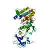
Mass: 614.202 Da / Num. of mol.: 1 / Source method: obtained synthetically / Formula: C30H40ClN7O3S
#3: Water ChemComp-HOH / water


Mass: 18.015 Da / Num. of mol.: 258 / Source method: isolated from a natural source / Formula: H2O

-
Experimental details

-
Experiment

ExperimentMethod: X-RAY DIFFRACTION / Number of used crystals: 1

-
Sample preparation

CrystalDensity Matthews: 2.27 Å3/Da / Density % sol: 45.87 %
Crystal growTemperature: 277 K / Method: vapor diffusion, sitting drop / pH: 8
Details: 0.1M SPG, 30.0% PEG 1K, pH 8.0, VAPOR DIFFUSION, SITTING DROP, temperature 277K

-
Data collection

DiffractionMean temperature: 100 K
Diffraction sourceSource: ROTATING ANODE / Type: RIGAKU FR-E SUPERBRIGHT / Wavelength: 1.54178 Å
DetectorType: RIGAKU RAXIS IV / Detector: IMAGE PLATE / Date: Jun 29, 2009
RadiationProtocol: SINGLE WAVELENGTH / Monochromatic (M) / Laue (L): M / Scattering type: x-ray
Radiation wavelengthWavelength: 1.54178 Å / Relative weight: 1
ReflectionRedundancy: 4.8 % / Av σ(I) over netI: 8 / Number: 160311 / Rsym value: 0.084 / D res high: 1.84 Å / D res low: 27.969 Å / Num. obs: 33290 / % possible obs: 98
Diffraction reflection shell
Highest resolution (Å)Lowest resolution (Å)% possible obs (%)IDRmerge(I) obsRsym valueRedundancy
5.8227.9799.110.0310.0314.6
4.115.8210010.0290.0294.8
3.364.1199.910.0410.0414.9
2.913.3699.310.0650.0654.9
2.62.9199.210.0920.0924.9
2.382.698.910.1390.1394.9
2.22.389810.1970.1974.9
2.062.297.210.2870.2874.8
1.942.0696.510.4210.4214.8
1.841.9495.910.7210.7214.7
ReflectionResolution: 1.84→28.93 Å / Num. all: 33969 / Num. obs: 33290 / % possible obs: 98 % / Redundancy: 4.8 % / Biso Wilson estimate: 24.6 Å2 / Rmerge(I) obs: 0.084 / Rsym value: 0.084 / Net I/σ(I): 12.5
Reflection shell

Diffraction-ID: 1

Resolution (Å)Redundancy (%)Rmerge(I) obsMean I/σ(I) obsNum. measured allNum. unique allRsym value% possible all
1.84-1.944.70.7211.12210847010.72195.9
1.94-2.064.80.4211.82153745070.42196.5
2.06-2.24.80.2872.72053642560.28797.2
2.2-2.384.90.1973.91930239760.19798
2.38-2.64.90.1395.51808737190.13998.9
2.6-2.914.90.0928.11650133910.09299.2
2.91-3.364.90.06510.81459729840.06599.3
3.36-4.114.90.04116.51265325930.04199.9
4.11-5.824.80.02922.6972920150.029100
5.82-27.9694.60.03114.7526111480.03199.1

-
Phasing

PhasingMethod: molecular replacement
Phasing MRRfactor: 48.63 / Model details: Phaser MODE: MR_AUTO
Highest resolutionLowest resolution
Rotation3.5 Å27.97 Å
Translation3.5 Å27.97 Å

-
Processing

Software
NameVersionClassificationNB
SCALA3.3.9data scaling
PHASER2.1.4phasing
REFMACrefinement
PDB_EXTRACT3.1data extraction
CrystalCleardata collection
MOSFLMdata reduction
RefinementMethod to determine structure: MOLECULAR REPLACEMENT
Starting model: PDB entry 3BKB
Resolution: 1.84→27.969 Å / Cor.coef. Fo:Fc: 0.954 / Cor.coef. Fo:Fc free: 0.928 / WRfactor Rfree: 0.2209 / WRfactor Rwork: 0.1714 / Occupancy max: 1 / Occupancy min: 0.4 / FOM work R set: 0.8755 / SU B: 5.361 / SU ML: 0.089 / SU R Cruickshank DPI: 0.1411 / SU Rfree: 0.1406 / Cross valid method: THROUGHOUT / σ(F): 0 / ESU R: 0.141 / ESU R Free: 0.141 / Stereochemistry target values: MAXIMUM LIKELIHOOD / Details: HYDROGENS HAVE BEEN ADDED IN THE RIDING POSITIONS
RfactorNum. reflection% reflectionSelection details
Rfree0.2379 1688 5.1 %RANDOM
Rwork0.1859 ---
all0.1885 33944 --
obs0.1885 33285 98.06 %-
Solvent computationIon probe radii: 0.8 Å / Shrinkage radii: 0.8 Å / VDW probe radii: 1.2 Å / Solvent model: MASK
Displacement parametersBiso max: 277.15 Å2 / Biso mean: 32.6003 Å2 / Biso min: 6.18 Å2
Baniso -1Baniso -2Baniso -3
1-0.22 Å20 Å2-0.42 Å2
2--0.35 Å20 Å2
3----0.66 Å2
Refinement stepCycle: LAST / Resolution: 1.84→27.969 Å
ProteinNucleic acidLigandSolventTotal
Num. atoms2787 0 42 258 3087
Refine LS restraints
Refine-IDTypeDev idealDev ideal targetNumber
X-RAY DIFFRACTIONr_bond_refined_d0.0150.022921
X-RAY DIFFRACTIONr_bond_other_d0.0010.021977
X-RAY DIFFRACTIONr_angle_refined_deg1.7261.9793977
X-RAY DIFFRACTIONr_angle_other_deg1.0553.0024775
X-RAY DIFFRACTIONr_dihedral_angle_1_deg6.0915358
X-RAY DIFFRACTIONr_dihedral_angle_2_deg32.06723.548124
X-RAY DIFFRACTIONr_dihedral_angle_3_deg14.72115464
X-RAY DIFFRACTIONr_dihedral_angle_4_deg16.9021519
X-RAY DIFFRACTIONr_chiral_restr0.1050.2440
X-RAY DIFFRACTIONr_gen_planes_refined0.0080.0213234
X-RAY DIFFRACTIONr_gen_planes_other0.0010.02595
LS refinement shellResolution: 1.84→1.888 Å / Total num. of bins used: 20
RfactorNum. reflection% reflection
Rfree0.376 99 -
Rwork0.362 2145 -
all-2244 -
obs--95.82 %
Refinement TLS params.

Method: refined / Refine-ID: X-RAY DIFFRACTION

IDL112)L122)L132)L222)L232)L332)S11 (Å °)S12 (Å °)S13 (Å °)S21 (Å °)S22 (Å °)S23 (Å °)S31 (Å °)S32 (Å °)S33 (Å °)T112)T122)T132)T222)T232)T332)Origin x (Å)Origin y (Å)Origin z (Å)
13.6253-0.06880.06273.25750.08065.06920.09110.1601-0.1153-0.2729-0.0576-0.3782-0.00251.1684-0.03360.18580.01560.00540.4385-0.02630.146416.912413.2191-7.5667
20.1536-0.32510.27151.2811-0.17135.0780.01660.07230.0309-0.32810.0536-0.0086-0.22320.1558-0.07020.1451-0.0904-0.01640.11710.03340.10582.722214.569918.9041
31.99760.4130.53481.84780.16931.1622-0.05770.1608-0.0244-0.0470.1181-0.01870.05020.049-0.06030.0117-0.00540.00940.0171-0.00110.03159.33274.327839.7784
Refinement TLS group
IDRefine-IDRefine TLS-IDAuth asym-IDAuth seq-ID
1X-RAY DIFFRACTION1A447 - 550
2X-RAY DIFFRACTION2A551 - 654
3X-RAY DIFFRACTION3A655 - 822

+
About Yorodumi

-
News

-
Feb 9, 2022. New format data for meta-information of EMDB entries

New format data for meta-information of EMDB entries

  • Version 3 of the EMDB header file is now the official format.
  • The previous official version 1.9 will be removed from the archive.

Related info.:EMDB header

External links:wwPDB to switch to version 3 of the EMDB data model

-
Aug 12, 2020. Covid-19 info

Covid-19 info

URL: https://pdbjlvh1.pdbj.org/emnavi/covid19.php

New page: Covid-19 featured information page in EM Navigator.

Related info.:Covid-19 info / Mar 5, 2020. Novel coronavirus structure data

+
Mar 5, 2020. Novel coronavirus structure data

Novel coronavirus structure data

Related info.:Yorodumi Speices / Aug 12, 2020. Covid-19 info

External links:COVID-19 featured content - PDBj / Molecule of the Month (242):Coronavirus Proteases

+
Jan 31, 2019. EMDB accession codes are about to change! (news from PDBe EMDB page)

EMDB accession codes are about to change! (news from PDBe EMDB page)

  • The allocation of 4 digits for EMDB accession codes will soon come to an end. Whilst these codes will remain in use, new EMDB accession codes will include an additional digit and will expand incrementally as the available range of codes is exhausted. The current 4-digit format prefixed with “EMD-” (i.e. EMD-XXXX) will advance to a 5-digit format (i.e. EMD-XXXXX), and so on. It is currently estimated that the 4-digit codes will be depleted around Spring 2019, at which point the 5-digit format will come into force.
  • The EM Navigator/Yorodumi systems omit the EMD- prefix.

Related info.:Q: What is EMD? / ID/Accession-code notation in Yorodumi/EM Navigator

External links:EMDB Accession Codes are Changing Soon! / Contact to PDBj

+
Jul 12, 2017. Major update of PDB

Major update of PDB

  • wwPDB released updated PDB data conforming to the new PDBx/mmCIF dictionary.
  • This is a major update changing the version number from 4 to 5, and with Remediation, in which all the entries are updated.
  • In this update, many items about electron microscopy experimental information are reorganized (e.g. em_software).
  • Now, EM Navigator and Yorodumi are based on the updated data.

External links:wwPDB Remediation / Enriched Model Files Conforming to OneDep Data Standards Now Available in the PDB FTP Archive

-
Yorodumi

Thousand views of thousand structures

  • Yorodumi is a browser for structure data from EMDB, PDB, SASBDB, etc.
  • This page is also the successor to EM Navigator detail page, and also detail information page/front-end page for Omokage search.
  • The word "yorodu" (or yorozu) is an old Japanese word meaning "ten thousand". "mi" (miru) is to see.

Related info.:EMDB / PDB / SASBDB / Comparison of 3 databanks / Yorodumi Search / Aug 31, 2016. New EM Navigator & Yorodumi / Yorodumi Papers / Jmol/JSmol / Function and homology information / Changes in new EM Navigator and Yorodumi

Read more