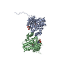
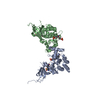
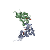
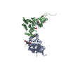
Entry Database : PDB / ID : 4e5wTitle JAK1 kinase (JH1 domain) in complex with compound 26 Tyrosine-protein kinase JAK1 Keywords / / / / Function / homology Function Domain/homology Component
/ / / / / / / / / / / / / / / / / / / / / / / / / / / / / / / / / / / / / / / / / / / / / / / / / / / / / / / / / / / / / / / / / / / / / / / / / / / / / / / / / / / / / / / / / / / / / / / / / / / / / / / / / / / / / / / / / / / / / / / / / / / / / / Biological species Homo sapiens (human)Method / / / / Resolution : 1.86 Å Authors Murray, J.M. Journal : J.Med.Chem. / Year : 2012Title : Identification of Imidazo-Pyrrolopyridines as Novel and Potent JAK1 Inhibitors.Authors: Kulagowski, J.J. / Blair, W. / Bull, R.J. / Chang, C. / Deshmukh, G. / Dyke, H.J. / Eigenbrot, C. / Ghilardi, N. / Gibbons, P. / Harrison, T.K. / Hewitt, P.R. / Liimatta, M. / Hurley, C.A. / ... Authors : Kulagowski, J.J. / Blair, W. / Bull, R.J. / Chang, C. / Deshmukh, G. / Dyke, H.J. / Eigenbrot, C. / Ghilardi, N. / Gibbons, P. / Harrison, T.K. / Hewitt, P.R. / Liimatta, M. / Hurley, C.A. / Johnson, A. / Johnson, T. / Kenny, J.R. / Bir Kohli, P. / Maxey, R.J. / Mendonca, R. / Mortara, K. / Murray, J. / Narukulla, R. / Shia, S. / Steffek, M. / Ubhayakar, S. / Ultsch, M. / van Abbema, A. / Ward, S.I. / Waszkowycz, B. / Zak, M. History Deposition Mar 14, 2012 Deposition site / Processing site Revision 1.0 May 30, 2012 Provider / Type Revision 1.1 Jul 11, 2012 Group Revision 1.2 Nov 6, 2024 Group Data collection / Database references ... Data collection / Database references / Derived calculations / Structure summary Category chem_comp_atom / chem_comp_bond ... chem_comp_atom / chem_comp_bond / database_2 / pdbx_entry_details / pdbx_modification_feature / struct_conn / struct_ref_seq_dif / struct_site Item _database_2.pdbx_DOI / _database_2.pdbx_database_accession ... _database_2.pdbx_DOI / _database_2.pdbx_database_accession / _struct_conn.pdbx_leaving_atom_flag / _struct_ref_seq_dif.details / _struct_site.pdbx_auth_asym_id / _struct_site.pdbx_auth_comp_id / _struct_site.pdbx_auth_seq_id
Show all Show less
 Movie
Movie Controller


 Open data
 Basic information
 Components
 Keywords
 Function and homology information
 Homo sapiens (human)
 X-RAY DIFFRACTION /
  Authors
 Citation
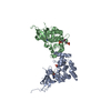
 Structure visualization
 Downloads & links
 Other downloads
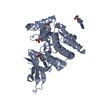


 PDBj
PDBj


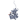
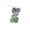
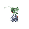
 Assembly
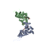

 Spodoptera frugiperda (fall armyworm)
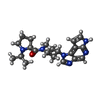
 Mass: 380.487 Da / Num. of mol.: 2 / Source method: obtained synthetically / Formula: C21H28N6O
 Mass: 106.120 Da / Num. of mol.: 1 / Source method: obtained synthetically / Formula: C4H10O3
 Mass: 18.015 Da / Num. of mol.: 421 / Source method: isolated from a natural source / Formula: H2O
 Sample preparation
 / Beamline: X06DA / Wavelength: 1 / Wavelength: 1 Å
 Processing