 Movie
Movie Controller

[English] 日本語
 Yorodumi
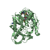
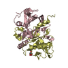
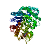
- PDB-4e2b: High resolution crystal structure of the old yellow enzyme from T... -
+ Open data

- Basic information
| Entry | Database: PDB / ID: 4e2b | ||||||
|---|---|---|---|---|---|---|---|
| Title | High resolution crystal structure of the old yellow enzyme from Trypanosoma cruzi | ||||||
 Components Components | Old Yellow Enzyme | ||||||
 Keywords Keywords | OXIDOREDUCTASE / TIM-barrel fold | ||||||
| Function / homology |  Function and homology information Function and homology information | ||||||
| Biological species |  | ||||||
| Method |  X-RAY DIFFRACTION / SYNCHROTRON / MOLECULAR REPLACEMENT / Resolution: 1.269 Å X-RAY DIFFRACTION / SYNCHROTRON / MOLECULAR REPLACEMENT / Resolution: 1.269 Å | ||||||
 Authors Authors | Murakami, M.T. / Rodrigues, N.C. / Gava, L.M. / Canduri, F. / Oliva, G. / Barbosa, L.R.S. / Borgers, J.C. | ||||||
 Citation Citation | Journal: To be Published Title: High resolution crystal structure and in solution studies of the old yellow enzyme from Trypanosoma cruzi: Insights into oligomerization, enzyme dynamics and specificity Authors: Murakami, M.T. / Rodrigues, N.C. / Gava, L.M. / Canduri, F. / Oliva, G. / Barbosa, L.R.S. / Borgers, J.C. | ||||||
| History |
|
- Structure visualization
| Structure viewer | Molecule: MolmilJmol/JSmol |
|---|
- Downloads & links
-Download
| PDBx/mmCIF format | 4e2b.cif.gz | 179.8 KB | Display | PDBx/mmCIF format |
|---|---|---|---|---|
| PDB format | pdb4e2b.ent.gz | 140.5 KB | Display | PDB format |
| PDBx/mmJSON format | 4e2b.json.gz | Tree view | PDBx/mmJSON format | |
| Others |  Other downloads Other downloads |
-Validation report
| Arichive directory | https://data.pdbj.org/pub/pdb/validation_reports/e2/4e2bftp://data.pdbj.org/pub/pdb/validation_reports/e2/4e2b | HTTPS FTP |
|---|
-Related structure data
| Related structure data |  3atyS S: Starting model for refinement |
|---|---|
| Similar structure data |


-Links
 PDBj
PDBj- Assembly
| Deposited unit | 
| ||||||||
|---|---|---|---|---|---|---|---|---|---|
| 1 |
| ||||||||
| Unit cell |
|
-Components
| #1: Protein | Mass: 42557.129 Da / Num. of mol.: 1 Source method: isolated from a genetically manipulated source Source: (gene. exp.)  | ||||
|---|---|---|---|---|---|
| #2: Chemical | ChemComp-FMN /   Mass: 456.344 Da / Num. of mol.: 1 / Source method: obtained synthetically / Formula: C17H21N4O9P Mass: 456.344 Da / Num. of mol.: 1 / Source method: obtained synthetically / Formula: C17H21N4O9P | ||||
| #3: Chemical |   Mass: 92.094 Da / Num. of mol.: 3 / Source method: obtained synthetically / Formula: C3H8O3 Mass: 92.094 Da / Num. of mol.: 3 / Source method: obtained synthetically / Formula: C3H8O3#4: Chemical |   Mass: 106.120 Da / Num. of mol.: 3 / Source method: obtained synthetically / Formula: C4H10O3 Mass: 106.120 Da / Num. of mol.: 3 / Source method: obtained synthetically / Formula: C4H10O3#5: Water | ChemComp-HOH / |  Mass: 18.015 Da / Num. of mol.: 363 / Source method: isolated from a natural source / Formula: H2O Mass: 18.015 Da / Num. of mol.: 363 / Source method: isolated from a natural source / Formula: H2O |
-Experimental details
-Experiment
| Experiment | Method: X-RAY DIFFRACTION / Number of used crystals: 1 |
|---|
- Sample preparation
| Crystal | Density Matthews: 1.97 Å3/Da / Density % sol: 37.57 % |
|---|---|
| Crystal grow | Temperature: 292 K / Method: vapor diffusion, hanging drop / pH: 8 Details: 28% (w/v) polyethylene glycol 1500 and 0.3 M ammonium fluoride, pH 8.0, VAPOR DIFFUSION, HANGING DROP, temperature 292K |
-Data collection
| Diffraction | Mean temperature: 100 K |
|---|---|
| Diffraction source | Source: SYNCHROTRON / Site: LNLS  / Beamline: W01B-MX2 / Wavelength: 1.46 Å / Beamline: W01B-MX2 / Wavelength: 1.46 Å |
| Detector | Type: MARMOSAIC 225 mm CCD / Detector: CCD / Date: Oct 10, 2011 |
| Radiation | Monochromator: Si(111) double-crystal / Protocol: SINGLE WAVELENGTH / Monochromatic (M) / Laue (L): M / Scattering type: x-ray |
| Radiation wavelength | Wavelength: 1.46 Å / Relative weight: 1 |
| Reflection | Resolution: 1.269→30 Å / Num. all: 105031 / Num. obs: 87922 / % possible obs: 98.3 % / Observed criterion σ(F): 1 / Observed criterion σ(I): 2 / Redundancy: 4.9 % / Rmerge(I) obs: 0.047 |
| Reflection shell | Resolution: 1.269→1.32 Å / Redundancy: 4.6 % / Rmerge(I) obs: 0.522 / % possible all: 99.9 |
- Processing
| Software |
| ||||||||||||||||||||||||||||||||||||||||||||||||||||||||||||
|---|---|---|---|---|---|---|---|---|---|---|---|---|---|---|---|---|---|---|---|---|---|---|---|---|---|---|---|---|---|---|---|---|---|---|---|---|---|---|---|---|---|---|---|---|---|---|---|---|---|---|---|---|---|---|---|---|---|---|---|---|---|
| Refinement | Method to determine structure: MOLECULAR REPLACEMENT Starting model: PDB ENTRY 3ATY Resolution: 1.269→22.48 Å / Cor.coef. Fo:Fc: 0.975 / Cor.coef. Fo:Fc free: 0.967 / SU B: 0.96 / SU ML: 0.018 / Cross valid method: THROUGHOUT / ESU R: 0.01 / ESU R Free: 0.01 / Stereochemistry target values: MAXIMUM LIKELIHOOD
| ||||||||||||||||||||||||||||||||||||||||||||||||||||||||||||
| Solvent computation | Ion probe radii: 0.8 Å / Shrinkage radii: 0.8 Å / VDW probe radii: 1.2 Å / Solvent model: MASK | ||||||||||||||||||||||||||||||||||||||||||||||||||||||||||||
| Displacement parameters | Biso mean: 14.677 Å2
| ||||||||||||||||||||||||||||||||||||||||||||||||||||||||||||
| Refinement step | Cycle: LAST / Resolution: 1.269→22.48 Å
| ||||||||||||||||||||||||||||||||||||||||||||||||||||||||||||
| Refine LS restraints |
| ||||||||||||||||||||||||||||||||||||||||||||||||||||||||||||
| LS refinement shell | Resolution: 1.269→1.302 Å / Total num. of bins used: 20
|
