#79 - Jul 2006 Amyloid-beta Precursor Protein similarity (1)
-
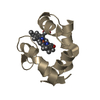
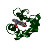
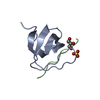
Assembly
Deposited unit
A: Major prion protein B: Major prion protein C: Major prion protein D: Major prion protein E: Major prion protein F: Major prion protein G: Major prion protein H: Major prion protein I: Major prion protein J: Major prion protein K: Major prion protein L: Major prion protein
Mass: 18.015 Da / Num. of mol.: 67 / Source method: isolated from a natural source / Formula: H2O
Compound details
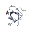
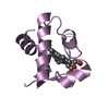
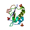
FRAGMENT OF HUMAN PRION PROTEIN FROM HELIX 2 AND 3
Has protein modification
Y
Source details
SEGMENTS CORRESPONDING TO RESIDUES 177-182 AND 211-216 OF HUMAN PRION WAS LINKED BY DISULFIDE BOND ...SEGMENTS CORRESPONDING TO RESIDUES 177-182 AND 211-216 OF HUMAN PRION WAS LINKED BY DISULFIDE BOND BETWEEN 179 AND 214 DURING SYNTHESIS
-
Experimental details
-
Experiment
Experiment
Method: X-RAY DIFFRACTION / Number of used crystals: 1
-
Sample preparation
Crystal
Density Matthews: 1.99 Å3/Da / Density % sol: 38.04 %
Crystal grow
Temperature: 298 K / Method: vapor diffusion, hanging drop / pH: 5.5 Details: 100mM Bis Tris pH 5.5, 25% PEG 3350, vapor diffusion, hanging drop, temperature 298K
Protocol: SINGLE WAVELENGTH / Monochromatic (M) / Laue (L): M / Scattering type: x-ray
Radiation wavelength
Wavelength: 1.5418 Å / Relative weight: 1
Reflection
Resolution: 2.028→30.873 Å / Num. all: 4685 / Num. obs: 4685 / % possible obs: 98.6 % / Redundancy: 5.7 % / Rsym value: 0.064 / Net I/σ(I): 20.1
Reflection shell
Diffraction-ID: 1
Resolution (Å)
Redundancy (%)
Rmerge(I) obs
Mean I/σ(I) obs
Num. measured all
Num. unique all
Rsym value
% possible all
2.028-2.14
3.8
0.196
3.9
2312
605
0.196
90.7
2.14-2.27
6
0.174
4.4
3806
637
0.174
99.9
2.27-2.42
6.1
0.14
5.5
3669
606
0.14
100
2.42-2.62
6.2
0.106
7.2
3469
564
0.106
100
2.62-2.87
6.1
0.093
7.9
3208
522
0.093
100
2.87-3.21
6.1
0.058
12.5
2925
480
0.058
100
3.21-3.7
6.1
0.044
15.4
2586
426
0.044
100
3.7-4.53
5.9
0.038
17.4
2185
372
0.038
99.9
4.53-6.41
5.7
0.037
15.3
1652
289
0.037
99.8
6.41-41.217
5
0.038
16.1
914
184
0.038
98.4
-
Phasing
Phasing
Method: molecular replacement
Phasing MR
Model details: Phaser MODE: MR_AUTO
Highest resolution
Lowest resolution
Rotation
2.03 Å
26.83 Å
Translation
2.03 Å
26.83 Å
-
Processing
Software
Name
Version
Classification
NB
SCALA
3.3.16
datascaling
PHASER
2.1.4
phasing
REFMAC
refinement
PDB_EXTRACT
3.1
dataextraction
XSCALE
datascaling
Refinement
Resolution: 2.03→23.3 Å / Cor.coef. Fo:Fc: 0.944 / Cor.coef. Fo:Fc free: 0.905 / SU B: 5.172 / SU ML: 0.131 / Cross valid method: THROUGHOUT / σ(F): 0 / ESU R: 0.234 / ESU R Free: 0.202 / Stereochemistry target values: MAXIMUM LIKELIHOOD Details: HYDROGENS HAVE BEEN ADDED IN THE RIDING POSITIONS U VALUES: WITH TLS ADDED
Rfactor
Num. reflection
% reflection
Selection details
Rfree
0.253
212
4.6 %
RANDOM
Rwork
0.187
-
-
-
obs
0.19
4649
98.3 %
-
Solvent computation
Ion probe radii: 0.8 Å / Shrinkage radii: 0.8 Å / VDW probe radii: 1.4 Å / Solvent model: MASK
Displacement parameters
Biso mean: 18.35 Å2
Baniso -1
Baniso -2
Baniso -3
1-
0.39 Å2
0 Å2
0 Å2
2-
-
0.07 Å2
0 Å2
3-
-
-
-0.45 Å2
Refinement step
Cycle: LAST / Resolution: 2.03→23.3 Å
Protein
Nucleic acid
Ligand
Solvent
Total
Num. atoms
576
0
0
67
643
Refine LS restraints
Refine-ID
Type
Dev ideal
Dev ideal target
Number
X-RAY DIFFRACTION
r_bond_refined_d
0.012
0.021
594
X-RAY DIFFRACTION
r_bond_other_d
0
0.02
334
X-RAY DIFFRACTION
r_angle_refined_deg
1.225
1.945
800
X-RAY DIFFRACTION
r_angle_other_deg
0.649
3
849
X-RAY DIFFRACTION
r_dihedral_angle_1_deg
10.208
5
63
X-RAY DIFFRACTION
r_dihedral_angle_2_deg
42.904
27.813
32
X-RAY DIFFRACTION
r_dihedral_angle_3_deg
17.085
15
111
X-RAY DIFFRACTION
r_dihedral_angle_4_deg
X-RAY DIFFRACTION
r_chiral_restr
0.081
0.2
98
X-RAY DIFFRACTION
r_gen_planes_refined
0.01
0.02
624
X-RAY DIFFRACTION
r_gen_planes_other
0.003
0.02
76
X-RAY DIFFRACTION
r_nbd_refined
X-RAY DIFFRACTION
r_nbd_other
X-RAY DIFFRACTION
r_nbtor_refined
X-RAY DIFFRACTION
r_nbtor_other
X-RAY DIFFRACTION
r_xyhbond_nbd_refined
X-RAY DIFFRACTION
r_xyhbond_nbd_other
X-RAY DIFFRACTION
r_metal_ion_refined
X-RAY DIFFRACTION
r_metal_ion_other
X-RAY DIFFRACTION
r_symmetry_vdw_refined
X-RAY DIFFRACTION
r_symmetry_vdw_other
X-RAY DIFFRACTION
r_symmetry_hbond_refined
X-RAY DIFFRACTION
r_symmetry_hbond_other
X-RAY DIFFRACTION
r_symmetry_metal_ion_refined
X-RAY DIFFRACTION
r_symmetry_metal_ion_other
X-RAY DIFFRACTION
r_mcbond_it
1.534
1.5
368
X-RAY DIFFRACTION
r_mcbond_other
0.412
1.5
134
X-RAY DIFFRACTION
r_mcangle_it
2.607
2
602
X-RAY DIFFRACTION
r_scbond_it
4.282
3
226
X-RAY DIFFRACTION
r_scangle_it
6.29
4.5
197
X-RAY DIFFRACTION
r_rigid_bond_restr
X-RAY DIFFRACTION
r_sphericity_free
X-RAY DIFFRACTION
r_sphericity_bonded
LS refinement shell
Resolution: 2.03→2.08 Å / Total num. of bins used: 20
Rfactor
Num. reflection
% reflection
Rfree
0.35
14
-
Rwork
0.205
275
-
obs
-
-
81.18 %
Refinement TLS params.
Method: refined / Origin x: -12.5408 Å / Origin y: 1.9013 Å / Origin z: 8.8177 Å
11
12
13
21
22
23
31
32
33
T
0.073 Å2
0.0483 Å2
0.0413 Å2
-
0.0573 Å2
0.015 Å2
-
-
0.0245 Å2
L
3.966 °2
1.2601 °2
3.8912 °2
-
2.7367 °2
0.905 °2
-
-
5.5905 °2
S
0.0837 Å °
-0.071 Å °
-0.0777 Å °
0.1232 Å °
0.2043 Å °
-0.038 Å °
-0.2999 Å °
-0.3194 Å °
-0.288 Å °
+
About Yorodumi
-
News
-
Feb 9, 2022. New format data for meta-information of EMDB entries
New format data for meta-information of EMDB entries
Version 3 of the EMDB header file is now the official format.
The previous official version 1.9 will be removed from the archive.
In the structure databanks used in Yorodumi, some data are registered as the other names, "COVID-19 virus" and "2019-nCoV". Here are the details of the virus and the list of structure data.
Jan 31, 2019. EMDB accession codes are about to change! (news from PDBe EMDB page)
EMDB accession codes are about to change! (news from PDBe EMDB page)
The allocation of 4 digits for EMDB accession codes will soon come to an end. Whilst these codes will remain in use, new EMDB accession codes will include an additional digit and will expand incrementally as the available range of codes is exhausted. The current 4-digit format prefixed with “EMD-” (i.e. EMD-XXXX) will advance to a 5-digit format (i.e. EMD-XXXXX), and so on. It is currently estimated that the 4-digit codes will be depleted around Spring 2019, at which point the 5-digit format will come into force.
The EM Navigator/Yorodumi systems omit the EMD- prefix.
Related info.:Q: What is EMD? / ID/Accession-code notation in Yorodumi/EM Navigator
Yorodumi is a browser for structure data from EMDB, PDB, SASBDB, etc.
This page is also the successor to EM Navigator detail page, and also detail information page/front-end page for Omokage search.
The word "yorodu" (or yorozu) is an old Japanese word meaning "ten thousand". "mi" (miru) is to see.
Related info.:EMDB / PDB / SASBDB / Comparison of 3 databanks / Yorodumi Search / Aug 31, 2016. New EM Navigator & Yorodumi / Yorodumi Papers / Jmol/JSmol / Function and homology information / Changes in new EM Navigator and Yorodumi
 Movie
Movie Controller


 Open data
 Basic information
 Components
 Keywords
 Function and homology information
 Homo sapiens (human)
 X-RAY DIFFRACTION /
  Authors
 Citation
 Structure visualization
 Downloads & links
 Other downloads


 PDBj
PDBj


 Assembly
 Mass: 18.015 Da / Num. of mol.: 67 / Source method: isolated from a natural source / Formula: H2O
 Sample preparation
 Processing