- PDB-4dum: Co-crystal structure of eIF4E with inhibitor -
+
Open data
ID or keywords:
Loading...
-
Basic information
Entry
Database: PDB / ID: 4dum
Title
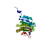
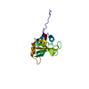
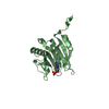
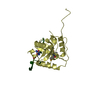
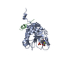
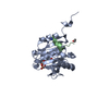
Co-crystal structure of eIF4E with inhibitor
Components
Eukaryotic translation initiation factor 4E
Keywords
TRANSLATION / CAP-binding protein / translation initiation factor / m7GTP
Function / homology
Function and homology information
eukaryotic initiation factor 4G binding / Activation of the mRNA upon binding of the cap-binding complex and eIFs, and subsequent binding to 43S / regulation of translation at postsynapse, modulating synaptic transmission / RNA cap binding / eukaryotic translation initiation factor 4F complex / Z-decay: degradation of maternal mRNAs by zygotically expressed factors / chromatoid body / Deadenylation of mRNA / mRNA cap binding / Transport of the SLBP independent Mature mRNA ...eukaryotic initiation factor 4G binding / Activation of the mRNA upon binding of the cap-binding complex and eIFs, and subsequent binding to 43S / regulation of translation at postsynapse, modulating synaptic transmission / RNA cap binding / eukaryotic translation initiation factor 4F complex / Z-decay: degradation of maternal mRNAs by zygotically expressed factors / chromatoid body / Deadenylation of mRNA / mRNA cap binding / Transport of the SLBP independent Mature mRNA / Transport of the SLBP Dependant Mature mRNA / RNA 7-methylguanosine cap binding / Transport of Mature mRNA Derived from an Intronless Transcript / M-decay: degradation of maternal mRNAs by maternally stored factors / RISC complex / negative regulation of neuron differentiation / stem cell population maintenance / Ribosomal scanning and start codon recognition / Translation initiation complex formation / mTORC1-mediated signalling / cellular response to dexamethasone stimulus / GTP hydrolysis and joining of the 60S ribosomal subunit / L13a-mediated translational silencing of Ceruloplasmin expression / mRNA export from nucleus / behavioral fear response / translation initiation factor activity / positive regulation of mitotic cell cycle / P-body / translational initiation / G1/S transition of mitotic cell cycle / ISG15 antiviral mechanism / neuron differentiation / cytoplasmic stress granule / cytoplasmic ribonucleoprotein granule / regulation of translation / DNA-binding transcription factor binding / negative regulation of translation / nuclear speck / postsynapse / perinuclear region of cytoplasm / glutamatergic synapse / enzyme binding / RNA binding / extracellular exosome / nucleus / cytoplasm / cytosol Similarity search - Function
Mass: 18.015 Da / Num. of mol.: 14 / Source method: isolated from a natural source / Formula: H2O
-
Experimental details
-
Experiment
Experiment
Method: X-RAY DIFFRACTION / Number of used crystals: 1
-
Sample preparation
Crystal
Density Matthews: 2.51 Å3/Da / Density % sol: 51.04 %
Crystal grow
Temperature: 289 K / Method: vapor diffusion, sitting drop Details: The purified protein which contained 100 uM m7-GTP was then concentrated to about 7 mg/mL in 20 mM Hepes, pH7.6, 100 mM KCl, 1mM DTT, 0.1 mM EDTA for crystallization. The m7-GTP-bound eIF4e ...Details: The purified protein which contained 100 uM m7-GTP was then concentrated to about 7 mg/mL in 20 mM Hepes, pH7.6, 100 mM KCl, 1mM DTT, 0.1 mM EDTA for crystallization. The m7-GTP-bound eIF4e protein was crystallized with 1:1 ratio of protein solution to reservoir solution of 17-20% PEG-3350 and 0.1-0.4M Na formate, VAPOR DIFFUSION, SITTING DROP, temperature 289K
Protocol: SINGLE WAVELENGTH / Monochromatic (M) / Laue (L): M / Scattering type: x-ray
Radiation wavelength
Wavelength: 1.54 Å / Relative weight: 1
Reflection
Resolution: 2.95→62.75 Å / Num. all: 18074 / Num. obs: 6282 / % possible obs: 98.9 % / Observed criterion σ(I): 2.9 / Redundancy: 2.9 % / Biso Wilson estimate: 58.7 Å2 / Rmerge(I) obs: 0.116 / Net I/σ(I): 9.5
Reflection shell
Resolution: 2.95→3.11 Å / Rmerge(I) obs: 0.324 / Mean I/σ(I) obs: 2.3 / Num. unique all: 765 / % possible all: 97.5
-
Processing
Software
Name
Version
Classification
StructureStudio
datacollection
MOLREP
phasing
REFMAC
5.5.0109
refinement
MOSFLM
datareduction
SCALA
datascaling
Refinement
Method to determine structure: MOLECULAR REPLACEMENT / Resolution: 2.95→38.236 Å / Cor.coef. Fo:Fc: 0.918 / Cor.coef. Fo:Fc free: 0.852 / SU B: 16.91 / SU ML: 0.317 / Cross valid method: THROUGHOUT / ESU R Free: 0.425 / Stereochemistry target values: MAXIMUM LIKELIHOOD / Details: HYDROGENS HAVE BEEN ADDED IN THE RIDING POSITIONS
Rfactor
Num. reflection
% reflection
Selection details
Rfree
0.2711
292
4.7 %
RANDOM
Rwork
0.20176
-
-
-
obs
0.20497
5969
98.46 %
-
Solvent computation
Ion probe radii: 0.8 Å / Shrinkage radii: 0.8 Å / VDW probe radii: 1.4 Å / Solvent model: MASK
Displacement parameters
Biso mean: 35.343 Å2
Baniso -1
Baniso -2
Baniso -3
1-
-0.08 Å2
0 Å2
0 Å2
2-
-
1.51 Å2
0 Å2
3-
-
-
-1.43 Å2
Refinement step
Cycle: LAST / Resolution: 2.95→38.236 Å
Protein
Nucleic acid
Ligand
Solvent
Total
Num. atoms
1547
0
40
14
1601
Refine LS restraints
Refine-ID
Type
Dev ideal
Dev ideal target
Number
X-RAY DIFFRACTION
r_bond_refined_d
0.014
0.022
1641
X-RAY DIFFRACTION
r_angle_refined_deg
1.506
1.953
2222
X-RAY DIFFRACTION
r_dihedral_angle_1_deg
6.718
5
189
X-RAY DIFFRACTION
r_dihedral_angle_2_deg
38.487
23.929
84
X-RAY DIFFRACTION
r_dihedral_angle_3_deg
19.904
15
284
X-RAY DIFFRACTION
r_dihedral_angle_4_deg
19.663
15
12
X-RAY DIFFRACTION
r_chiral_restr
0.1
0.2
228
X-RAY DIFFRACTION
r_gen_planes_refined
0.006
0.021
1255
X-RAY DIFFRACTION
r_mcbond_it
0.818
1.5
935
X-RAY DIFFRACTION
r_mcangle_it
1.581
2
1512
X-RAY DIFFRACTION
r_scbond_it
1.835
3
706
X-RAY DIFFRACTION
r_scangle_it
3.215
4.5
708
LS refinement shell
Resolution: 2.95→3.027 Å / Total num. of bins used: 20
Rfactor
Num. reflection
% reflection
Rfree
0.401
19
-
Rwork
0.276
401
-
obs
-
-
97.22 %
+
About Yorodumi
-
News
-
Feb 9, 2022. New format data for meta-information of EMDB entries
New format data for meta-information of EMDB entries
Version 3 of the EMDB header file is now the official format.
The previous official version 1.9 will be removed from the archive.
In the structure databanks used in Yorodumi, some data are registered as the other names, "COVID-19 virus" and "2019-nCoV". Here are the details of the virus and the list of structure data.
Jan 31, 2019. EMDB accession codes are about to change! (news from PDBe EMDB page)
EMDB accession codes are about to change! (news from PDBe EMDB page)
The allocation of 4 digits for EMDB accession codes will soon come to an end. Whilst these codes will remain in use, new EMDB accession codes will include an additional digit and will expand incrementally as the available range of codes is exhausted. The current 4-digit format prefixed with “EMD-” (i.e. EMD-XXXX) will advance to a 5-digit format (i.e. EMD-XXXXX), and so on. It is currently estimated that the 4-digit codes will be depleted around Spring 2019, at which point the 5-digit format will come into force.
The EM Navigator/Yorodumi systems omit the EMD- prefix.
Related info.:Q: What is EMD? / ID/Accession-code notation in Yorodumi/EM Navigator
Yorodumi is a browser for structure data from EMDB, PDB, SASBDB, etc.
This page is also the successor to EM Navigator detail page, and also detail information page/front-end page for Omokage search.
The word "yorodu" (or yorozu) is an old Japanese word meaning "ten thousand". "mi" (miru) is to see.
Related info.:EMDB / PDB / SASBDB / Comparison of 3 databanks / Yorodumi Search / Aug 31, 2016. New EM Navigator & Yorodumi / Yorodumi Papers / Jmol/JSmol / Function and homology information / Changes in new EM Navigator and Yorodumi
 Movie
Movie Controller


 Open data
 Basic information
 Components
 Keywords
 Function and homology information
 Homo sapiens (human)
 X-RAY DIFFRACTION /
  Authors
 Citation
 Structure visualization
 Downloads & links
 Other downloads
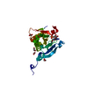


 PDBj
PDBj


 Assembly
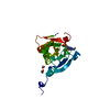


 Mass: 475.822 Da / Num. of mol.: 1 / Source method: obtained synthetically / Formula: C20H19ClN5O5P
 Mass: 62.068 Da / Num. of mol.: 2 / Source method: obtained synthetically / Formula: C2H6O2
 Mass: 18.015 Da / Num. of mol.: 14 / Source method: isolated from a natural source / Formula: H2O
 Sample preparation
 Processing