 Movie
Movie Controller

[English] 日本語
 Yorodumi
- PDB-4dp7: The 1.08 Angstrom crystal structure of oxidized (CuII) poplar pla... -
+ Open data

- Basic information
| Entry | Database: PDB / ID: 4dp7 | ||||||
|---|---|---|---|---|---|---|---|
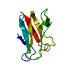
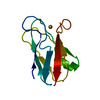
| Title | The 1.08 Angstrom crystal structure of oxidized (CuII) poplar plastocyanin A at pH 4.0 | ||||||
 Components Components | Plastocyanin A, chloroplastic | ||||||
 Keywords Keywords | ELECTRON TRANSPORT / membrane / thylakoid / transit peptide / plastocyanin-like domain / copper-binding | ||||||
| Function / homology |  Function and homology information Function and homology informationchloroplast thylakoid lumen / chloroplast thylakoid membrane / electron transfer activity / copper ion binding Similarity search - Function | ||||||
| Biological species |  | ||||||
| Method |  X-RAY DIFFRACTION / SYNCHROTRON / MOLECULAR REPLACEMENT / Resolution: 1.08 Å X-RAY DIFFRACTION / SYNCHROTRON / MOLECULAR REPLACEMENT / Resolution: 1.08 Å | ||||||
 Authors Authors | Kachalova, G.S. / Shosheva, A.H. / Bourenkov, G.P. / Donchev, A.A. / Dimitrov, M.I. / Bartunik, H.D. | ||||||
 Citation Citation | Journal: J.Inorg.Biochem. / Year: 2012 Title: Structural comparison of the poplar plastocyanin isoforms PCa and PCb sheds new light on the role of the copper site geometry in interactions with redox partners in oxygenic photosynthesis. Authors: Kachalova, G.S. / Shosheva, A.C. / Bourenkov, G.P. / Donchev, A.A. / Dimitrov, M.I. / Bartunik, H.D. | ||||||
| History |
|
- Structure visualization
| Structure viewer | Molecule: MolmilJmol/JSmol |
|---|
- Downloads & links
-Download
| PDBx/mmCIF format | 4dp7.cif.gz | 63.6 KB | Display | PDBx/mmCIF format |
|---|---|---|---|---|
| PDB format | pdb4dp7.ent.gz | 47.2 KB | Display | PDB format |
| PDBx/mmJSON format | 4dp7.json.gz | Tree view | PDBx/mmJSON format | |
| Others |  Other downloads Other downloads |
-Validation report
| Arichive directory | https://data.pdbj.org/pub/pdb/validation_reports/dp/4dp7ftp://data.pdbj.org/pub/pdb/validation_reports/dp/4dp7 | HTTPS FTP |
|---|
-Related structure data
| Related structure data | 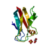 4dp0C  4dp1C  4dp2C  4dp4C 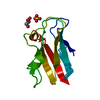 4dp5C  4dp6C  4dp8C  4dp9C  4dpaC  4dpbC  4dpcC  1plcS S: Starting model for refinement C: citing same article ( |
|---|---|
| Similar structure data |


-Links
 PDBj
PDBj- Assembly
| Deposited unit | 
| ||||||||
|---|---|---|---|---|---|---|---|---|---|
| 1 |
| ||||||||
| Unit cell |
|
-Components
| #1: Protein | Mass: 10493.607 Da / Num. of mol.: 1 / Fragment: UNP residues 70-168 / Source method: isolated from a natural source / Source: (natural) |
|---|---|
| #2: Chemical | ChemComp-CU /   Mass: 63.546 Da / Num. of mol.: 1 / Source method: obtained synthetically / Formula: Cu Mass: 63.546 Da / Num. of mol.: 1 / Source method: obtained synthetically / Formula: Cu |
| #3: Chemical | ChemComp-SO4 /   Mass: 96.063 Da / Num. of mol.: 1 / Source method: obtained synthetically / Formula: SO4 Mass: 96.063 Da / Num. of mol.: 1 / Source method: obtained synthetically / Formula: SO4 |
| #4: Water | ChemComp-HOH /  Mass: 18.015 Da / Num. of mol.: 177 / Source method: isolated from a natural source / Formula: H2O Mass: 18.015 Da / Num. of mol.: 177 / Source method: isolated from a natural source / Formula: H2O |
-Experimental details
-Experiment
| Experiment | Method: X-RAY DIFFRACTION / Number of used crystals: 1 |
|---|
- Sample preparation
| Crystal | Density Matthews: 1.83 Å3/Da / Density % sol: 32.75 % |
|---|---|
| Crystal grow | Temperature: 277 K / Method: vapor diffusion, hanging drop / pH: 4 Details: 2.6 M ammonium sulfate, 0.1 M sodium phosphate buffer, pH 6.0, VAPOR DIFFUSION, HANGING DROP, temperature 277K |
-Data collection
| Diffraction | Mean temperature: 100 K |
|---|---|
| Diffraction source | Source: SYNCHROTRON / Site: MPG/DESY, HAMBURG  / Beamline: BW6 / Wavelength: 1.05 Å / Beamline: BW6 / Wavelength: 1.05 Å |
| Detector | Type: MAR CCD 165 mm / Detector: CCD / Date: Mar 3, 2002 / Details: MIRRORS |
| Radiation | Monochromator: double crystal Si(111) / Protocol: SINGLE WAVELENGTH / Monochromatic (M) / Laue (L): M / Scattering type: x-ray |
| Radiation wavelength | Wavelength: 1.05 Å / Relative weight: 1 |
| Reflection | Resolution: 1.08→35.99 Å / Num. all: 32914 / Num. obs: 32728 / % possible obs: 99.4 % / Observed criterion σ(I): 2 / Redundancy: 4.2 % / Biso Wilson estimate: 7.98 Å2 / Rmerge(I) obs: 0.062 / Net I/σ(I): 18.3 |
| Reflection shell | Resolution: 1.08→1.1 Å / Redundancy: 3.9 % / Rmerge(I) obs: 0.401 / Mean I/σ(I) obs: 2.3 / Num. unique all: 1585 / % possible all: 98.7 |
- Processing
| Software |
| |||||||||||||||||||||||||||||||||||||||||||||||||||||||
|---|---|---|---|---|---|---|---|---|---|---|---|---|---|---|---|---|---|---|---|---|---|---|---|---|---|---|---|---|---|---|---|---|---|---|---|---|---|---|---|---|---|---|---|---|---|---|---|---|---|---|---|---|---|---|---|---|
| Refinement | Method to determine structure: MOLECULAR REPLACEMENT Starting model: PDB ENTRY 1PLC Resolution: 1.08→35.99 Å / Cor.coef. Fo:Fc: 0.979 / Cor.coef. Fo:Fc free: 0.973 / SU B: 0.721 / SU ML: 0.016 / Isotropic thermal model: ANISOTROPIC / Cross valid method: THROUGHOUT / ESU R: 0.031 / ESU R Free: 0.029 / Stereochemistry target values: MAXIMUM LIKELIHOOD
| |||||||||||||||||||||||||||||||||||||||||||||||||||||||
| Solvent computation | Ion probe radii: 0.8 Å / Shrinkage radii: 0.8 Å / VDW probe radii: 1.4 Å / Solvent model: MASK | |||||||||||||||||||||||||||||||||||||||||||||||||||||||
| Displacement parameters | Biso mean: 12.676 Å2
| |||||||||||||||||||||||||||||||||||||||||||||||||||||||
| Refinement step | Cycle: LAST / Resolution: 1.08→35.99 Å
| |||||||||||||||||||||||||||||||||||||||||||||||||||||||
| Refine LS restraints |
| |||||||||||||||||||||||||||||||||||||||||||||||||||||||
| LS refinement shell | Resolution: 1.08→1.108 Å / Total num. of bins used: 20
|
