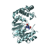
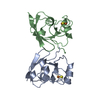
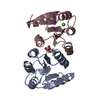
- PDB-4dn6: Crystal structure of a putative pilus assembly protein (cpaE) fro... -
+
Open data
ID or keywords:
Loading...
-
Basic information
Entry
Database: PDB / ID: 4dn6
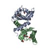
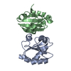
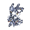
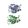
Title
Crystal structure of a putative pilus assembly protein (cpaE) from Burkholderia thailandensis E264 at 2.80 A resolution
Components
Putative pilus assembly protein CpaE
Keywords
SIGNALING PROTEIN / Response regulator receiver domain / CHEY-related protein / signal transduction / Structural Genomics / Joint Center for Structural Genomics / JCSG / Protein Structure Initiative / PSI-BIOLOGY
Function / homology
Function and homology information
negative regulation of cell division / phosphorelay signal transduction system / cytoplasmic side of plasma membrane / ATP hydrolysis activity / ATP binding / cytosol Similarity search - Function
Mass: 18.015 Da / Num. of mol.: 16 / Source method: isolated from a natural source / Formula: H2O
Has protein modification
Y
Sequence details
THE CONSTRUCT WAS EXPRESSED WITH AN N-TERMINAL PURIFICATION TAG MGSDKIHHHHHHENLYFQG FOLLOWED BY ...THE CONSTRUCT WAS EXPRESSED WITH AN N-TERMINAL PURIFICATION TAG MGSDKIHHHHHHENLYFQG FOLLOWED BY RESIDUES 1-127 OF THE FULL LENGTH PROTEIN.
-
Experimental details
-
Experiment
Experiment
Method: X-RAY DIFFRACTION / Number of used crystals: 1
-
Sample preparation
Crystal
Density Matthews: 2.37 Å3/Da / Density % sol: 48.03 %
Crystal grow
Temperature: 277 K / Method: vapor diffusion, sitting drop / pH: 7 Details: 10.0% PEG-6000, 0.1M HEPES pH 7.0, NANODROP, VAPOR DIFFUSION, SITTING DROP, temperature 277K
Resolution: 2.8→29.57 Å / Num. obs: 8094 / % possible obs: 98.6 % / Observed criterion σ(I): -3 / Redundancy: 6.89 % / Biso Wilson estimate: 93.904 Å2 / Rmerge(I) obs: 0.057 / Net I/σ(I): 15.71
Reflection shell
Resolution (Å)
Rmerge(I) obs
Mean I/σ(I) obs
Num. measured obs
Num. unique obs
Diffraction-ID
% possible all
2.8-2.9
0.795
1.58
5759
1484
1
98.5
2.9-3.02
0.474
2.7
5841
1495
1
99.4
3.02-3.15
0.32
4
5206
1390
1
99.6
3.15-3.32
0.225
6.1
5329
1491
1
99.3
3.32-3.52
0.109
10.6
5827
1437
1
99.6
3.52-3.79
0.098
14.3
5273
1395
1
94.3
3.79-4.17
0.056
20.4
5843
1489
1
99.5
4.17-4.77
0.042
27.6
5124
1429
1
98.3
4.77-5.98
0.038
30.5
5796
1479
1
99.8
5.98-29.57
0.025
38.5
5740
1506
1
98
-
Phasing
Phasing
Method: MAD
-
Processing
Software
Name
Version
Classification
NB
MolProbity
3beta29
modelbuilding
PDB_EXTRACT
3.1
dataextraction
SHELX
phasing
SHARP
phasing
XSCALE
December6, 2010
datascaling
BUSTER-TNT
2.10.0
refinement
XDS
datareduction
SHELXD
phasing
BUSTER
2.10.0
refinement
Refinement
Method to determine structure: MAD / Resolution: 2.8→29.57 Å / Cor.coef. Fo:Fc: 0.9418 / Cor.coef. Fo:Fc free: 0.9314 / Occupancy max: 1 / Occupancy min: 0.5 / Cross valid method: THROUGHOUT / σ(F): 0 Details: 1. A MET-INHIBITION PROTOCOL WAS USED FOR SELENOMETHIONINE INCORPORATION DURING PROTEIN EXPRESSION. THE OCCUPANCY OF THE SE ATOMS IN THE MSE RESIDUES WAS REDUCED TO 0.75 FOR THE REDUCED ...Details: 1. A MET-INHIBITION PROTOCOL WAS USED FOR SELENOMETHIONINE INCORPORATION DURING PROTEIN EXPRESSION. THE OCCUPANCY OF THE SE ATOMS IN THE MSE RESIDUES WAS REDUCED TO 0.75 FOR THE REDUCED SCATTERING POWER DUE TO PARTIAL S-MET INCORPORATION. 2. ATOM RECORD CONTAINS SUM OF TLS AND RESIDUAL B FACTORS. ANISOU RECORD CONTAINS SUM OF TLS AND RESIDUAL U FACTORS. 3. WATERS WERE EXCLUDED FROM AUTOMATIC TLS ASSIGNMENT. 4. NCS RESTRAINTS WERE APPLIED USING BUSTER'S LSSR RESTRAINT REPRESENTATION (-AUTONCS). 5. MAD EXPERIMENTAL PHASES IN THE FORM OF HENDRICKSON-LATTMAN COEFFICIENTS WERE USED AS RESTRAINTS DURING REFINEMENT.
In the structure databanks used in Yorodumi, some data are registered as the other names, "COVID-19 virus" and "2019-nCoV". Here are the details of the virus and the list of structure data.
Jan 31, 2019. EMDB accession codes are about to change! (news from PDBe EMDB page)
EMDB accession codes are about to change! (news from PDBe EMDB page)
The allocation of 4 digits for EMDB accession codes will soon come to an end. Whilst these codes will remain in use, new EMDB accession codes will include an additional digit and will expand incrementally as the available range of codes is exhausted. The current 4-digit format prefixed with “EMD-” (i.e. EMD-XXXX) will advance to a 5-digit format (i.e. EMD-XXXXX), and so on. It is currently estimated that the 4-digit codes will be depleted around Spring 2019, at which point the 5-digit format will come into force.
The EM Navigator/Yorodumi systems omit the EMD- prefix.
Related info.:Q: What is EMD? / ID/Accession-code notation in Yorodumi/EM Navigator
Yorodumi is a browser for structure data from EMDB, PDB, SASBDB, etc.
This page is also the successor to EM Navigator detail page, and also detail information page/front-end page for Omokage search.
The word "yorodu" (or yorozu) is an old Japanese word meaning "ten thousand". "mi" (miru) is to see.
Related info.:EMDB / PDB / SASBDB / Comparison of 3 databanks / Yorodumi Search / Aug 31, 2016. New EM Navigator & Yorodumi / Yorodumi Papers / Jmol/JSmol / Function and homology information / Changes in new EM Navigator and Yorodumi
 Movie
Movie Controller

 Yorodumi
 Open data
 Basic information
 Components
 Keywords
 Function and homology information
 Burkholderia thailandensis (bacteria)
 X-RAY DIFFRACTION /
  Authors
 Citation
 Structure visualization
 Downloads & links
 Other downloads


 PDBj
PDBj

 Assembly
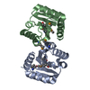
 Mass: 18.015 Da / Num. of mol.: 16 / Source method: isolated from a natural source / Formula: H2O
 Sample preparation
 / Beamline: BL11-1 / Wavelength: 0.97895,0.91837
 Processing