 Movie
Movie Controller

[English] 日本語
 Yorodumi
- PDB-4dl8: Crystal structure of Trypanosoma brucei dUTPase with dUMP, planar... -
+ Open data

- Basic information
| Entry | Database: PDB / ID: 4dl8 | ||||||
|---|---|---|---|---|---|---|---|
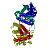
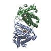
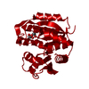
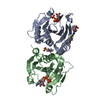
| Title | Crystal structure of Trypanosoma brucei dUTPase with dUMP, planar [AlF3-OPO3] transition state analogue, Mg2+, and Na+ | ||||||
 Components Components | Deoxyuridine triphosphatase | ||||||
 Keywords Keywords | HYDROLASE / all alpha NTP pyrophosphohydrolase / all alpha NTP pyrophosphatase | ||||||
| Function / homology |  Function and homology information Function and homology informationdUTP diphosphatase / dUTP diphosphatase activity / nucleoplasm / metal ion binding / cytosol Similarity search - Function | ||||||
| Biological species |  | ||||||
| Method |  X-RAY DIFFRACTION / SYNCHROTRON / REFINED 4DKB / Resolution: 1.698 Å X-RAY DIFFRACTION / SYNCHROTRON / REFINED 4DKB / Resolution: 1.698 Å | ||||||
 Authors Authors | Hemsworth, G.R. / Gonzalez-Pacanowska, D. / Wilson, K.S. | ||||||
 Citation Citation | Journal: Biochem.J. / Year: 2013 Title: On the catalytic mechanism of dimeric dUTPases. Authors: Hemsworth, G.R. / Gonzalez-Pacanowska, D. / Wilson, K.S. | ||||||
| History |
|
- Structure visualization
| Structure viewer | Molecule: MolmilJmol/JSmol |
|---|
- Downloads & links
-Download
| PDBx/mmCIF format | 4dl8.cif.gz | 67.2 KB | Display | PDBx/mmCIF format |
|---|---|---|---|---|
| PDB format | pdb4dl8.ent.gz | 46 KB | Display | PDB format |
| PDBx/mmJSON format | 4dl8.json.gz | Tree view | PDBx/mmJSON format | |
| Others |  Other downloads Other downloads |
-Validation report
| Arichive directory | https://data.pdbj.org/pub/pdb/validation_reports/dl/4dl8ftp://data.pdbj.org/pub/pdb/validation_reports/dl/4dl8 | HTTPS FTP |
|---|
-Related structure data
| Related structure data | 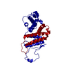 4dk2C  4dk4C 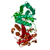 4dkbSC  4dlcC C: citing same article ( S: Starting model for refinement |
|---|---|
| Similar structure data |


-Links
 PDBj
PDBj


- Assembly
| Deposited unit | 
| ||||||||
|---|---|---|---|---|---|---|---|---|---|
| 1 | 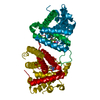
| ||||||||
| Unit cell |
|
-Components
-Protein , 1 types, 1 molecules A
| #1: Protein | Mass: 32204.271 Da / Num. of mol.: 1 Source method: isolated from a genetically manipulated source Source: (gene. exp.)  |
|---|
-Non-polymers , 6 types, 172 molecules 


| #2: Chemical | ChemComp-UMP /  Mass: 308.182 Da / Num. of mol.: 1 / Source method: obtained synthetically / Formula: C9H13N2O8P Mass: 308.182 Da / Num. of mol.: 1 / Source method: obtained synthetically / Formula: C9H13N2O8P | ||||
|---|---|---|---|---|---|
| #3: Chemical | ChemComp-AF3 /  Mass: 83.977 Da / Num. of mol.: 1 / Source method: obtained synthetically / Formula: AlF3 Mass: 83.977 Da / Num. of mol.: 1 / Source method: obtained synthetically / Formula: AlF3 | ||||
| #4: Chemical | ChemComp-PO4 /  Mass: 94.971 Da / Num. of mol.: 1 / Source method: obtained synthetically / Formula: PO4 Mass: 94.971 Da / Num. of mol.: 1 / Source method: obtained synthetically / Formula: PO4 | ||||
| #5: Chemical |  Mass: 24.305 Da / Num. of mol.: 2 / Source method: obtained synthetically / Formula: Mg Mass: 24.305 Da / Num. of mol.: 2 / Source method: obtained synthetically / Formula: Mg#6: Chemical | ChemComp-NA / |  Mass: 22.990 Da / Num. of mol.: 1 / Source method: obtained synthetically / Formula: Na Mass: 22.990 Da / Num. of mol.: 1 / Source method: obtained synthetically / Formula: Na#7: Water | ChemComp-HOH / | Mass: 18.015 Da / Num. of mol.: 166 / Source method: isolated from a natural source / Formula: H2O |
-Experimental details
-Experiment
| Experiment | Method: X-RAY DIFFRACTION / Number of used crystals: 1 |
|---|
- Sample preparation
| Crystal | Density Matthews: 2.26 Å3/Da / Density % sol: 45.57 % |
|---|---|
| Crystal grow | Temperature: 290 K / Method: vapor diffusion, sitting drop / pH: 6.5 Details: 0.2 M sodium sulfate, 0.1 M Bis-Tris propane, pH 6.5, 20% w/v PEG3350, VAPOR DIFFUSION, SITTING DROP, temperature 290K |
-Data collection
| Diffraction | Mean temperature: 100 K | ||||||||||||||||||||||||||||||||||||||||||||||||||||||||||||||||||||||||||||||||||||||||
|---|---|---|---|---|---|---|---|---|---|---|---|---|---|---|---|---|---|---|---|---|---|---|---|---|---|---|---|---|---|---|---|---|---|---|---|---|---|---|---|---|---|---|---|---|---|---|---|---|---|---|---|---|---|---|---|---|---|---|---|---|---|---|---|---|---|---|---|---|---|---|---|---|---|---|---|---|---|---|---|---|---|---|---|---|---|---|---|---|---|
| Diffraction source | Source: SYNCHROTRON / Site: Diamond  / Beamline: I04-1 / Wavelength: 0.917 Å / Beamline: I04-1 / Wavelength: 0.917 Å | ||||||||||||||||||||||||||||||||||||||||||||||||||||||||||||||||||||||||||||||||||||||||
| Detector | Type: DECTRIS PILATUS 2M / Detector: PIXEL / Date: Jan 28, 2011 | ||||||||||||||||||||||||||||||||||||||||||||||||||||||||||||||||||||||||||||||||||||||||
| Radiation | Monochromator: Si(111) / Protocol: SINGLE WAVELENGTH / Monochromatic (M) / Laue (L): M / Scattering type: x-ray | ||||||||||||||||||||||||||||||||||||||||||||||||||||||||||||||||||||||||||||||||||||||||
| Radiation wavelength | Wavelength: 0.917 Å / Relative weight: 1 | ||||||||||||||||||||||||||||||||||||||||||||||||||||||||||||||||||||||||||||||||||||||||
| Reflection | Resolution: 1.698→28.203 Å / Num. all: 33398 / Num. obs: 33398 / % possible obs: 99.7 % / Observed criterion σ(F): 1 / Observed criterion σ(I): 1 / Redundancy: 15.7 % / Biso Wilson estimate: 23.8 Å2 / Rmerge(I) obs: 0.078 / Rsym value: 0.078 / Net I/σ(I): 23.5 | ||||||||||||||||||||||||||||||||||||||||||||||||||||||||||||||||||||||||||||||||||||||||
| Reflection shell | Diffraction-ID: 1
|
- Processing
| Software |
| |||||||||||||||||||||||||||||||||||||||||||||||||||||||||||||||||||||||||||||||||||||
|---|---|---|---|---|---|---|---|---|---|---|---|---|---|---|---|---|---|---|---|---|---|---|---|---|---|---|---|---|---|---|---|---|---|---|---|---|---|---|---|---|---|---|---|---|---|---|---|---|---|---|---|---|---|---|---|---|---|---|---|---|---|---|---|---|---|---|---|---|---|---|---|---|---|---|---|---|---|---|---|---|---|---|---|---|---|---|
| Refinement | Method to determine structure: REFINED 4DKB Starting model: PDB ENTRY 4DKB Resolution: 1.698→28.2 Å / Cor.coef. Fo:Fc: 0.969 / Cor.coef. Fo:Fc free: 0.957 / WRfactor Rfree: 0.1759 / WRfactor Rwork: 0.1545 / Occupancy max: 1 / Occupancy min: 1 / FOM work R set: 0.8909 / SU B: 1.681 / SU ML: 0.055 / SU R Cruickshank DPI: 0.0828 / SU Rfree: 0.0837 / Cross valid method: THROUGHOUT / σ(F): 0 / ESU R: 0.083 / ESU R Free: 0.084 / Stereochemistry target values: MAXIMUM LIKELIHOOD Details: HYDROGENS HAVE BEEN ADDED IN THE RIDING POSITIONS U VALUES : REFINED INDIVIDUALLY
| |||||||||||||||||||||||||||||||||||||||||||||||||||||||||||||||||||||||||||||||||||||
| Solvent computation | Ion probe radii: 0.8 Å / Shrinkage radii: 0.8 Å / VDW probe radii: 1.4 Å / Solvent model: MASK | |||||||||||||||||||||||||||||||||||||||||||||||||||||||||||||||||||||||||||||||||||||
| Displacement parameters | Biso max: 424.2 Å2 / Biso mean: 25.9297 Å2 / Biso min: 8.23 Å2
| |||||||||||||||||||||||||||||||||||||||||||||||||||||||||||||||||||||||||||||||||||||
| Refinement step | Cycle: LAST / Resolution: 1.698→28.2 Å
| |||||||||||||||||||||||||||||||||||||||||||||||||||||||||||||||||||||||||||||||||||||
| Refine LS restraints |
| |||||||||||||||||||||||||||||||||||||||||||||||||||||||||||||||||||||||||||||||||||||
| LS refinement shell | Resolution: 1.698→1.742 Å / Total num. of bins used: 20
|
