 Movie
Movie Controller

[English] 日本語
 Yorodumi
- PDB-4dcz: Crystal structure of a domain from a mycoplasma genitalium termin... -
+ Open data

- Basic information
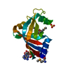
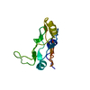
| Entry | Database: PDB / ID: 4dcz | ||||||
|---|---|---|---|---|---|---|---|
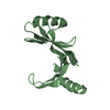
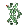
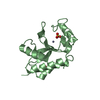
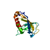
| Title | Crystal structure of a domain from a mycoplasma genitalium terminal organelle protein | ||||||
 Components Components | DnaJ-like protein MG200 | ||||||
 Keywords Keywords | UNKNOWN FUNCTION / DIMER / INTRA-DOMAIN SYMMETRY AXIS | ||||||
| Function / homology |  Function and homology information Function and homology information | ||||||
| Biological species |  Mycoplasma genitalium (bacteria) Mycoplasma genitalium (bacteria) | ||||||
| Method |  X-RAY DIFFRACTION / SYNCHROTRON / SAD / Resolution: 2.9 Å X-RAY DIFFRACTION / SYNCHROTRON / SAD / Resolution: 2.9 Å | ||||||
 Authors Authors | Calisto, B.M. / Martinelli, L. / Fita, I. | ||||||
 Citation Citation | Journal: Mol.Microbiol. / Year: 2012 Title: The EAGR box structure: a motif involved in mycoplasma motility. Authors: Calisto, B.M. / Broto, A. / Martinelli, L. / Querol, E. / Pinol, J. / Fita, I. | ||||||
| History |
|
- Structure visualization
| Structure viewer | Molecule: MolmilJmol/JSmol |
|---|
- Downloads & links
-Download
| PDBx/mmCIF format | 4dcz.cif.gz | 59.9 KB | Display | PDBx/mmCIF format |
|---|---|---|---|---|
| PDB format | pdb4dcz.ent.gz | 44.9 KB | Display | PDB format |
| PDBx/mmJSON format | 4dcz.json.gz | Tree view | PDBx/mmJSON format | |
| Others |  Other downloads Other downloads |
-Validation report
| Arichive directory | https://data.pdbj.org/pub/pdb/validation_reports/dc/4dczftp://data.pdbj.org/pub/pdb/validation_reports/dc/4dcz | HTTPS FTP |
|---|
-Related structure data
| Similar structure data |
|---|


-Links
 PDBj
PDBj

- Assembly
| Deposited unit | 
| ||||||||
|---|---|---|---|---|---|---|---|---|---|
| 1 |
| ||||||||
| 2 | 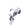
| ||||||||
| 3 | 
| ||||||||
| Unit cell |
| ||||||||
| Details | BIOMOLECULE: 1, 2, 3 SEE REMARK 350 FOR THE AUTHOR PROVIDED AND/OR PROGRAM GENERATED ASSEMBLY INFORMATION FOR THE STRUCTURE IN THIS ENTRY. THE REMARK MAY ALSO PROVIDE INFORMATION ON BURIED SURFACE AREA. COORDINATES FOR A COMPLETE MULTIMER REPRESENTING THE KNOWN BIOLOGICALLY SIGNIFICANT OLIGOMERIZATION STATE OF THE MOLECULE CAN BE GENERATED BY APPLYING BIOMT TRANSFORMATIONS GIVEN BELOW. BOTH NON-CRYSTALLOGRAPHIC AND CRYSTALLOGRAPHIC OPERATIONS ARE GIVEN. BIOMOLECULE: 1 AUTHOR DETERMINED BIOLOGICAL UNIT: DIMERIC APPLY THE FOLLOWING TO CHAINS: A, B BIOMT1 1 1.000000 0.000000 0.000000 0.00000 BIOMT2 1 0.000000 1.000000 0.000000 0.00000 BIOMT3 1 0.000000 0.000000 1.000000 0.00000 BIOMOLECULE: 2 SOFTWARE DETERMINED QUATERNARY STRUCTURE: MONOMERIC SOFTWARE USED: PISA APPLY THE FOLLOWING TO CHAINS: B BIOMT1 1 1.000000 0.000000 0.000000 0.00000 BIOMT2 1 0.000000 1.000000 0.000000 0.00000 BIOMT3 1 0.000000 0.000000 1.000000 0.00000 BIOMOLECULE: 3 SOFTWARE DETERMINED QUATERNARY STRUCTURE: MONOMERIC SOFTWARE USED: PISA APPLY THE FOLLOWING TO CHAINS: A BIOMT1 1 1.000000 0.000000 0.000000 0.00000 BIOMT2 1 0.000000 1.000000 0.000000 0.00000 BIOMT3 1 0.000000 0.000000 1.000000 0.00000 |
-Components
| #1: Protein | Mass: 10938.724 Da / Num. of mol.: 2 / Fragment: TO PROTEIN DOMAIN (UNP RESIDUES 124-207) Source method: isolated from a genetically manipulated source Source: (gene. exp.) Mycoplasma genitalium (bacteria) / Strain: G37 / Gene: MG200 / Plasmid: PET21D / Production host: |
|---|
-Experimental details
-Experiment
| Experiment | Method: X-RAY DIFFRACTION / Number of used crystals: 2 |
|---|
- Sample preparation
| Crystal | Density Matthews: 3.17 Å3/Da / Density % sol: 61.24 % |
|---|---|
| Crystal grow | Temperature: 293 K / Method: vapor diffusion, hanging drop / pH: 4.5 Details: 22% PEG 2000, 0.1 M SODIUM CITRATE, PH 4.5, VAPOR DIFFUSION, HANGING DROP, TEMPERATURE 293K |
-Data collection
| Diffraction |
| ||||||||||||||||||
|---|---|---|---|---|---|---|---|---|---|---|---|---|---|---|---|---|---|---|---|
| Diffraction source |
| ||||||||||||||||||
| Detector |
| ||||||||||||||||||
| Radiation |
| ||||||||||||||||||
| Radiation wavelength | Wavelength: 0.979 Å / Relative weight: 1 | ||||||||||||||||||
| Reflection | Resolution: 2.9→30 Å / Num. obs: 6450 / % possible obs: 74 % / Observed criterion σ(F): 0 / Observed criterion σ(I): -3 / Redundancy: 10.5 % / Biso Wilson estimate: 70 Å2 / Rmerge(I) obs: 0.067 / Rsym value: 0.066 / Net I/σ(I): 32.7 | ||||||||||||||||||
| Reflection shell | Resolution: 2.9→3 Å / Redundancy: 10 % / Rmerge(I) obs: 0.498 / Mean I/σ(I) obs: 5.2 / Rsym value: 0.393 / % possible all: 90 |

- Processing
| Software |
| |||||||||||||||||||||||||||||||||||||||||||||||||||||||||||||||||||||||||||||||||||||||||||||||||||||||||||||||||||||||||||||||||||||||||||||||||||||||||||||||||||||||||||||||
|---|---|---|---|---|---|---|---|---|---|---|---|---|---|---|---|---|---|---|---|---|---|---|---|---|---|---|---|---|---|---|---|---|---|---|---|---|---|---|---|---|---|---|---|---|---|---|---|---|---|---|---|---|---|---|---|---|---|---|---|---|---|---|---|---|---|---|---|---|---|---|---|---|---|---|---|---|---|---|---|---|---|---|---|---|---|---|---|---|---|---|---|---|---|---|---|---|---|---|---|---|---|---|---|---|---|---|---|---|---|---|---|---|---|---|---|---|---|---|---|---|---|---|---|---|---|---|---|---|---|---|---|---|---|---|---|---|---|---|---|---|---|---|---|---|---|---|---|---|---|---|---|---|---|---|---|---|---|---|---|---|---|---|---|---|---|---|---|---|---|---|---|---|---|---|---|---|
| Refinement | Method to determine structure: SAD / Resolution: 2.9→27.18 Å / Cor.coef. Fo:Fc: 0.936 / Cor.coef. Fo:Fc free: 0.882 / SU B: 25.518 / SU ML: 0.217 / Cross valid method: THROUGHOUT / σ(F): 0 / ESU R Free: 0.298 / Stereochemistry target values: MAXIMUM LIKELIHOOD / Details: HYDROGENS HAVE BEEN ADDED IN THE RIDING
| |||||||||||||||||||||||||||||||||||||||||||||||||||||||||||||||||||||||||||||||||||||||||||||||||||||||||||||||||||||||||||||||||||||||||||||||||||||||||||||||||||||||||||||||
| Solvent computation | Ion probe radii: 0.8 Å / Shrinkage radii: 0.8 Å / VDW probe radii: 1.4 Å / Solvent model: MASK | |||||||||||||||||||||||||||||||||||||||||||||||||||||||||||||||||||||||||||||||||||||||||||||||||||||||||||||||||||||||||||||||||||||||||||||||||||||||||||||||||||||||||||||||
| Displacement parameters | Biso mean: 62.51 Å2
| |||||||||||||||||||||||||||||||||||||||||||||||||||||||||||||||||||||||||||||||||||||||||||||||||||||||||||||||||||||||||||||||||||||||||||||||||||||||||||||||||||||||||||||||
| Refinement step | Cycle: LAST / Resolution: 2.9→27.18 Å
| |||||||||||||||||||||||||||||||||||||||||||||||||||||||||||||||||||||||||||||||||||||||||||||||||||||||||||||||||||||||||||||||||||||||||||||||||||||||||||||||||||||||||||||||
| Refine LS restraints |
| |||||||||||||||||||||||||||||||||||||||||||||||||||||||||||||||||||||||||||||||||||||||||||||||||||||||||||||||||||||||||||||||||||||||||||||||||||||||||||||||||||||||||||||||
| LS refinement shell | Resolution: 2.9→2.97 Å / Total num. of bins used: 20
| |||||||||||||||||||||||||||||||||||||||||||||||||||||||||||||||||||||||||||||||||||||||||||||||||||||||||||||||||||||||||||||||||||||||||||||||||||||||||||||||||||||||||||||||
| Refinement TLS params. | Method: refined / Refine-ID: X-RAY DIFFRACTION
| |||||||||||||||||||||||||||||||||||||||||||||||||||||||||||||||||||||||||||||||||||||||||||||||||||||||||||||||||||||||||||||||||||||||||||||||||||||||||||||||||||||||||||||||
| Refinement TLS group |
|
