regulation of translation by machinery localization / protein storage / structural constituent of cytoplasmic lattice / cytoplasmic lattice / cytoplasm organization / cortical granule / embryonic cleavage / intermediate filament cytoskeleton / epigenetic programming in the zygotic pronuclei / regulation of epidermal cell division ...regulation of translation by machinery localization / protein storage / structural constituent of cytoplasmic lattice / cytoplasmic lattice / cytoplasm organization / cortical granule / embryonic cleavage / intermediate filament cytoskeleton / epigenetic programming in the zygotic pronuclei / regulation of epidermal cell division / protein kinase C inhibitor activity / positive regulation of epidermal cell differentiation / keratinocyte development / keratinization / regulation of cell-cell adhesion / cAMP/PKA signal transduction / Regulation of localization of FOXO transcription factors / keratinocyte proliferation / phosphoserine residue binding / Activation of BAD and translocation to mitochondria / negative regulation of keratinocyte proliferation / establishment of skin barrier / negative regulation of protein localization to plasma membrane / Chk1/Chk2(Cds1) mediated inactivation of Cyclin B:Cdk1 complex / SARS-CoV-2 targets host intracellular signalling and regulatory pathways / negative regulation of protein kinase activity / negative regulation of stem cell proliferation / Chromatin modifying enzymes / SARS-CoV-1 targets host intracellular signalling and regulatory pathways / RHO GTPases activate PKNs / positive regulation of protein localization / cytoskeleton organization / tubulin binding / positive regulation of cell adhesion / protein sequestering activity / negative regulation of innate immune response / protein export from nucleus / TP53 Regulates Transcription of Genes Involved in G2 Cell Cycle Arrest / release of cytochrome c from mitochondria / positive regulation of protein export from nucleus / stem cell proliferation / Translocation of SLC2A4 (GLUT4) to the plasma membrane / TP53 Regulates Metabolic Genes / intrinsic apoptotic signaling pathway in response to DNA damage / intracellular protein localization / regulation of protein localization / positive regulation of cell growth / in utero embryonic development / regulation of cell cycle / cadherin binding / calcium ion binding / protein kinase binding / negative regulation of transcription by RNA polymerase II / signal transduction / extracellular space / extracellular exosome / identical protein binding / nucleus / cytosol / cytoplasm 類似検索 - 分子機能
 ムービー
ムービー コントローラー


 データを開く
 基本情報
基本情報 要素
要素 キーワード
キーワード 機能・相同性情報
 Homo sapiens (ヒト)
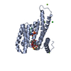
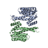
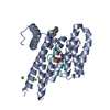
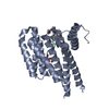
 X線回折 / 解像度: 2 Å
  データ登録者
 引用
引用 構造の表示
構造の表示 ダウンロードとリンク
 その他のダウンロード


 PDBj
PDBj


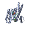
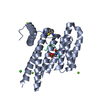
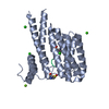
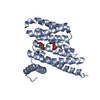
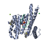
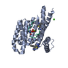
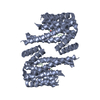
 集合体
集合体

 分子量: 18.015 Da / 分子数: 388 / 由来タイプ: 天然 / 式: H2O
 試料調製
試料調製 解析
解析