 Movie
Movie Controller


+ Open data

- Basic information
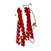
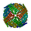
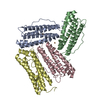
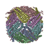
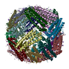
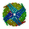
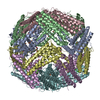
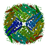
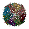
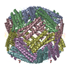
| Entry | Database: PDB / ID: 4das | ||||||
|---|---|---|---|---|---|---|---|
| Title | Crystal structure of Bullfrog M ferritin | ||||||
 Components Components | Ferritin, middle subunit | ||||||
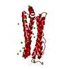
 Keywords Keywords | OXIDOREDUCTASE / iron storage protein / 24-subunit maxiferritin / four-helix bundle subunit / ferroxidase | ||||||
| Function / homology |  Function and homology information Function and homology informationferroxidase / ferroxidase activity / ferric iron binding / iron ion transport / ferrous iron binding / intracellular iron ion homeostasis / cytoplasm Similarity search - Function | ||||||
| Biological species | Rana catesbeiana (American bullfrog) | ||||||
| Method |  X-RAY DIFFRACTION / SYNCHROTRON / MOLECULAR REPLACEMENT / Resolution: 2.56 Å X-RAY DIFFRACTION / SYNCHROTRON / MOLECULAR REPLACEMENT / Resolution: 2.56 Å | ||||||
 Authors Authors | Bertini, I. / Lalli, D. / Mangani, S. / Pozzi, C. / Rosa, C. / Turano, P. | ||||||
 Citation Citation | Journal: J.Am.Chem.Soc. / Year: 2012 Title: Structural insights into the ferroxidase site of ferritins from higher eukaryotes. Authors: Bertini, I. / Lalli, D. / Mangani, S. / Pozzi, C. / Rosa, C. / Theil, E.C. / Turano, P. | ||||||
| History |
|
- Structure visualization
| Structure viewer | Molecule: MolmilJmol/JSmol |
|---|
- Downloads & links
-Download
| PDBx/mmCIF format | 4das.cif.gz | 841.9 KB | Display | PDBx/mmCIF format |
|---|---|---|---|---|
| PDB format | pdb4das.ent.gz | 706.2 KB | Display | PDB format |
| PDBx/mmJSON format | 4das.json.gz | Tree view | PDBx/mmJSON format | |
| Others |  Other downloads Other downloads |
-Validation report
| Summary document | 4das_validation.pdf.gz | 616.3 KB | Display | wwPDB validaton report |
|---|---|---|---|---|
| Full document | 4das_full_validation.pdf.gz | 683.2 KB | Display | |
| Data in XML | 4das_validation.xml.gz | 157.8 KB | Display | |
| Data in CIF | 4das_validation.cif.gz | 215.5 KB | Display | |
| Arichive directory | https://data.pdbj.org/pub/pdb/validation_reports/da/4dasftp://data.pdbj.org/pub/pdb/validation_reports/da/4das | HTTPS FTP |
-Related structure data
| Related structure data |  3rbcC 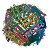 3re7C 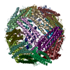 3rgdC 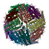 1mfrS C: citing same article ( S: Starting model for refinement |
|---|---|
| Similar structure data |


-Links
 PDBj
PDBj


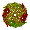
- Assembly
| Deposited unit | 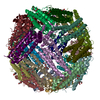
| ||||||||
|---|---|---|---|---|---|---|---|---|---|
| 1 |
| ||||||||
| Unit cell |
|
-Components
| #1: Protein | Mass: 20623.182 Da / Num. of mol.: 24 Source method: isolated from a genetically manipulated source Source: (gene. exp.) Rana catesbeiana (American bullfrog) / Plasmid: pET3a / Production host:  #2: Chemical | ChemComp-PGE /   Mass: 150.173 Da / Num. of mol.: 22 / Source method: obtained synthetically / Formula: C6H14O4 Mass: 150.173 Da / Num. of mol.: 22 / Source method: obtained synthetically / Formula: C6H14O4#3: Chemical | ChemComp-EDO /   Mass: 62.068 Da / Num. of mol.: 12 / Source method: obtained synthetically / Formula: C2H6O2 Mass: 62.068 Da / Num. of mol.: 12 / Source method: obtained synthetically / Formula: C2H6O2#4: Water | ChemComp-HOH / |  Mass: 18.015 Da / Num. of mol.: 1525 / Source method: isolated from a natural source / Formula: H2O Mass: 18.015 Da / Num. of mol.: 1525 / Source method: isolated from a natural source / Formula: H2O |
|---|
-Experimental details
-Experiment
| Experiment | Method: X-RAY DIFFRACTION / Number of used crystals: 1 |
|---|
- Sample preparation
| Crystal | Density Matthews: 4.19 Å3/Da / Density % sol: 70.66 % |
|---|---|
| Crystal grow | Temperature: 298 K / Method: vapor diffusion, sitting drop / pH: 7.5 Details: 3.0 M sodium formate, pH 7.5, VAPOR DIFFUSION, SITTING DROP, temperature 298.0K |
-Data collection
| Diffraction | Mean temperature: 100 K |
|---|---|
| Diffraction source | Source: SYNCHROTRON / Site: ESRF  / Beamline: ID23-1 / Wavelength: 0.97625 Å / Beamline: ID23-1 / Wavelength: 0.97625 Å |
| Detector | Type: ADSC QUANTUM 315r / Detector: CCD / Date: Jul 27, 2009 / Details: Toroidal Mirror |
| Radiation | Monochromator: Silicon (1 1 1) channel-cut / Protocol: SINGLE WAVELENGTH / Monochromatic (M) / Laue (L): M / Scattering type: x-ray |
| Radiation wavelength | Wavelength: 0.97625 Å / Relative weight: 1 |
| Reflection | Resolution: 2.56→100.5 Å / Num. all: 259578 / Num. obs: 259578 / % possible obs: 98 % / Observed criterion σ(I): 2 / Redundancy: 1.9 % / Biso Wilson estimate: 42.386 Å2 / Rmerge(I) obs: 0.115 / Net I/σ(I): 5.2 |
| Reflection shell | Resolution: 2.56→2.7 Å / Redundancy: 1.9 % / Rmerge(I) obs: 0.362 / Mean I/σ(I) obs: 1.7 / Num. unique all: 37996 / % possible all: 98.5 |
- Processing
| Software |
| |||||||||||||||||||||||||||||||||||||||||||||||||||||||||||||||||
|---|---|---|---|---|---|---|---|---|---|---|---|---|---|---|---|---|---|---|---|---|---|---|---|---|---|---|---|---|---|---|---|---|---|---|---|---|---|---|---|---|---|---|---|---|---|---|---|---|---|---|---|---|---|---|---|---|---|---|---|---|---|---|---|---|---|---|
| Refinement | Method to determine structure: MOLECULAR REPLACEMENT Starting model: PDB entry 1MFR Resolution: 2.56→79.51 Å / Cor.coef. Fo:Fc: 0.939 / Cor.coef. Fo:Fc free: 0.901 / SU B: 8.278 / SU ML: 0.175 / Cross valid method: THROUGHOUT / σ(I): 2 / ESU R: 0.288 / ESU R Free: 0.239 / Stereochemistry target values: MAXIMUM LIKELIHOOD
| |||||||||||||||||||||||||||||||||||||||||||||||||||||||||||||||||
| Solvent computation | Ion probe radii: 0.8 Å / Shrinkage radii: 0.8 Å / VDW probe radii: 1.4 Å / Solvent model: MASK | |||||||||||||||||||||||||||||||||||||||||||||||||||||||||||||||||
| Displacement parameters | Biso mean: 31.184 Å2
| |||||||||||||||||||||||||||||||||||||||||||||||||||||||||||||||||
| Refinement step | Cycle: LAST / Resolution: 2.56→79.51 Å
| |||||||||||||||||||||||||||||||||||||||||||||||||||||||||||||||||
| Refine LS restraints |
| |||||||||||||||||||||||||||||||||||||||||||||||||||||||||||||||||
| LS refinement shell | Resolution: 2.56→2.626 Å / Total num. of bins used: 20
|
