 Movie
Movie Controller


+ Open data

- Basic information
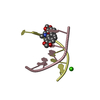
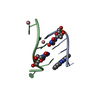
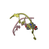
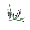
| Entry | Database: PDB / ID: 4d9y | ||||||||||||||||||
|---|---|---|---|---|---|---|---|---|---|---|---|---|---|---|---|---|---|---|---|
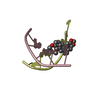
| Title | The crystal structure of Chelerythrine bound to DNA d(CGTACG) | ||||||||||||||||||
 Components Components | DNA (5'-D(* Keywords KeywordsDNA / DRUG-DNA COMPLEX / DOUBLE HELIX | Function / homology | Chem-CTI / DNA |  Function and homology information Function and homology informationMethod |  X-RAY DIFFRACTION / SYNCHROTRON / MOLECULAR REPLACEMENT / molecular replacement / Resolution: 2.1 Å X-RAY DIFFRACTION / SYNCHROTRON / MOLECULAR REPLACEMENT / molecular replacement / Resolution: 2.1 Å  Authors AuthorsFerraroni, M. / Bazzicalupi, C. / Gratteri, P. / Bilia, A.R. |  CitationJournal: To be Published CitationJournal: To be PublishedTitle: The crystal structure of Chelerythrine bound to DNA d(CGTACG) Authors: Ferraroni, M. / Bazzicalupi, C. / Gratteri, P. / Bilia, A.R. History |
|
- Structure visualization
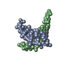
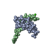
| Structure viewer | Molecule: MolmilJmol/JSmol |
|---|
- Downloads & links
-Download
| PDBx/mmCIF format | 4d9y.cif.gz | 24.2 KB | Display | PDBx/mmCIF format |
|---|---|---|---|---|
| PDB format | pdb4d9y.ent.gz | 14.5 KB | Display | PDB format |
| PDBx/mmJSON format | 4d9y.json.gz | Tree view | PDBx/mmJSON format | |
| Others |  Other downloads Other downloads |
-Validation report
| Arichive directory | https://data.pdbj.org/pub/pdb/validation_reports/d9/4d9yftp://data.pdbj.org/pub/pdb/validation_reports/d9/4d9y | HTTPS FTP |
|---|
-Related structure data
| Related structure data | 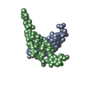 3np6S S: Starting model for refinement |
|---|---|
| Similar structure data |

-Links
 PDBj
PDBj

- Assembly
| Deposited unit | 
| ||||||||
|---|---|---|---|---|---|---|---|---|---|
| 1 | 
| ||||||||
| 2 | 
| ||||||||
| Unit cell |
|
-Components
| #1: DNA chain | Mass: 1809.217 Da / Num. of mol.: 4 / Source method: obtained synthetically / Details: synthetic DNA #2: Chemical | ChemComp-CTI / |   Mass: 348.372 Da / Num. of mol.: 1 / Source method: obtained synthetically / Formula: C21H18NO4 Mass: 348.372 Da / Num. of mol.: 1 / Source method: obtained synthetically / Formula: C21H18NO4#3: Chemical | ChemComp-CA / |   Mass: 40.078 Da / Num. of mol.: 1 / Source method: obtained synthetically / Formula: Ca Mass: 40.078 Da / Num. of mol.: 1 / Source method: obtained synthetically / Formula: Ca#4: Water | ChemComp-HOH / |  Mass: 18.015 Da / Num. of mol.: 11 / Source method: isolated from a natural source / Formula: H2O Mass: 18.015 Da / Num. of mol.: 11 / Source method: isolated from a natural source / Formula: H2O |
|---|
-Experimental details
-Experiment
| Experiment | Method: X-RAY DIFFRACTION / Number of used crystals: 1 |
|---|
- Sample preparation
| Crystal | Density Matthews: 2.19 Å3/Da / Density % sol: 43.89 % |
|---|---|
| Crystal grow | Temperature: 296 K / Method: vapor diffusion / pH: 6.5 Details: MPD, MgCl2, NaCl, KCL, spermine, pH 6.5, VAPOR DIFFUSION, temperature 296K |
-Data collection
| Diffraction | Mean temperature: 100 K | ||||||||||||||||||||||||||||||||||||||||||||||||||
|---|---|---|---|---|---|---|---|---|---|---|---|---|---|---|---|---|---|---|---|---|---|---|---|---|---|---|---|---|---|---|---|---|---|---|---|---|---|---|---|---|---|---|---|---|---|---|---|---|---|---|---|
| Diffraction source | Source: SYNCHROTRON / Site: ELETTRA  / Beamline: 5.2R / Wavelength: 1 Å / Beamline: 5.2R / Wavelength: 1 Å | ||||||||||||||||||||||||||||||||||||||||||||||||||
| Detector | Type: PILATUS 2M / Detector: CCD / Date: Mar 13, 2011 | ||||||||||||||||||||||||||||||||||||||||||||||||||
| Radiation | Monochromator: double-crystal Si(111) / Protocol: SINGLE WAVELENGTH / Monochromatic (M) / Laue (L): M / Scattering type: x-ray | ||||||||||||||||||||||||||||||||||||||||||||||||||
| Radiation wavelength | Wavelength: 1 Å / Relative weight: 1 | ||||||||||||||||||||||||||||||||||||||||||||||||||
| Reflection | Resolution: 2.1→30 Å / Num. obs: 4087 / % possible obs: 99 % / Observed criterion σ(I): -3 / Biso Wilson estimate: 46.83 Å2 / Rmerge(I) obs: 0.089 / Net I/σ(I): 13.39 | ||||||||||||||||||||||||||||||||||||||||||||||||||
| Reflection shell |
|
-Phasing
| Phasing | Method: molecular replacement | |||||||||
|---|---|---|---|---|---|---|---|---|---|---|
| Phasing MR |
|
- Processing
| Software |
| |||||||||||||||||||||||||||||||||
|---|---|---|---|---|---|---|---|---|---|---|---|---|---|---|---|---|---|---|---|---|---|---|---|---|---|---|---|---|---|---|---|---|---|---|
| Refinement | Method to determine structure: MOLECULAR REPLACEMENT Starting model: PDB ENTRY 3NP6 Resolution: 2.1→20 Å / Occupancy max: 1 / Occupancy min: 1 / Cross valid method: FREE R / σ(F): 0 / Stereochemistry target values: Engh & Huber Details: Crystal was merohedrally twinned with a fraction of 0.46 and twin law -h, -k, l
| |||||||||||||||||||||||||||||||||
| Displacement parameters | Biso mean: 54.1262 Å2 | |||||||||||||||||||||||||||||||||
| Refinement step | Cycle: LAST / Resolution: 2.1→20 Å
| |||||||||||||||||||||||||||||||||
| Refine LS restraints |
| |||||||||||||||||||||||||||||||||
| LS refinement shell | Resolution: 2.1→2.19 Å /
|
