 Movie
Movie Controller

[English] 日本語
 Yorodumi
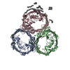
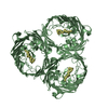
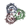
- PDB-4d64: Structure of porin Omp-Pst1 from P. stuartii; the crystallographi... -
+ Open data

- Basic information
| Entry | Database: PDB / ID: 4d64 | ||||||
|---|---|---|---|---|---|---|---|
| Title | Structure of porin Omp-Pst1 from P. stuartii; the crystallographic symmetry generates a dimer of trimers. | ||||||
 Components Components | PORIN 1 | ||||||
 Keywords Keywords | TRANSPORT PROTEIN / BACTERIAL JUNCTION / STERIC-ZIPPER / DIMER OF TRIMERS | ||||||
| Function / homology |  Function and homology information Function and homology informationporin activity / pore complex / cell outer membrane / monoatomic ion transmembrane transport Similarity search - Function | ||||||
| Biological species |  PROVIDENCIA STUARTII (bacteria) PROVIDENCIA STUARTII (bacteria) | ||||||
| Method |  X-RAY DIFFRACTION / SYNCHROTRON / MOLECULAR REPLACEMENT / Resolution: 3.2 Å X-RAY DIFFRACTION / SYNCHROTRON / MOLECULAR REPLACEMENT / Resolution: 3.2 Å | ||||||
 Authors Authors | Nasrallah, C. / Colletier, J.P. | ||||||
 Citation Citation | Journal: Proc. Natl. Acad. Sci. U.S.A. / Year: 2018 Title: Porin self-association enables cell-to-cell contact inProvidencia stuartiifloating communities. Authors: El-Khatib, M. / Nasrallah, C. / Lopes, J. / Tran, Q.T. / Tetreau, G. / Basbous, H. / Fenel, D. / Gallet, B. / Lethier, M. / Bolla, J.M. / Pages, J.M. / Vivaudou, M. / Weik, M. / ...Authors: El-Khatib, M. / Nasrallah, C. / Lopes, J. / Tran, Q.T. / Tetreau, G. / Basbous, H. / Fenel, D. / Gallet, B. / Lethier, M. / Bolla, J.M. / Pages, J.M. / Vivaudou, M. / Weik, M. / Winterhalter, M. / Colletier, J.P. #1: Journal: Plos One / Year: 2015 Title: Understanding Voltage Gating of Providencia Stuartii Porins at Atomic Level. Authors: Song, W. / Bajaj, H. / Nasrallah, C. / Jiang, H. / Winterhalter, M. / Colletier, J. / Xu, Y. | ||||||
| History |
|
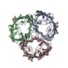
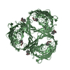
- Structure visualization
| Structure viewer | Molecule: MolmilJmol/JSmol |
|---|
- Downloads & links
-Download
| PDBx/mmCIF format | 4d64.cif.gz | 423.4 KB | Display | PDBx/mmCIF format |
|---|---|---|---|---|
| PDB format | pdb4d64.ent.gz | 352.9 KB | Display | PDB format |
| PDBx/mmJSON format | 4d64.json.gz | Tree view | PDBx/mmJSON format | |
| Others |  Other downloads Other downloads |
-Validation report
| Arichive directory | https://data.pdbj.org/pub/pdb/validation_reports/d6/4d64ftp://data.pdbj.org/pub/pdb/validation_reports/d6/4d64 | HTTPS FTP |
|---|
-Related structure data
| Related structure data |  4d65C  5n9hC  5n9iC  5nxnC  5nxrC  5nxuC  1opfS  4d5w 4d5x C: citing same article ( S: Starting model for refinement |
|---|---|
| Similar structure data |


-Links
 PDBj
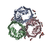
PDBj- Assembly
| Deposited unit | 
| ||||||||||||||||||||||||
|---|---|---|---|---|---|---|---|---|---|---|---|---|---|---|---|---|---|---|---|---|---|---|---|---|---|
| 1 |
| ||||||||||||||||||||||||
| Unit cell |
| ||||||||||||||||||||||||
| Noncrystallographic symmetry (NCS) | NCS domain:
NCS domain segments:
|
-Components
| #1: Protein | Mass: 39034.527 Da / Num. of mol.: 3 Source method: isolated from a genetically manipulated source Source: (gene. exp.) PROVIDENCIA STUARTII (bacteria) / Plasmid: PG_OMP-PST1 / Production host: #2: Chemical | ChemComp-CA / |   Mass: 40.078 Da / Num. of mol.: 1 / Source method: obtained synthetically / Formula: Ca Mass: 40.078 Da / Num. of mol.: 1 / Source method: obtained synthetically / Formula: Ca#3: Water | ChemComp-HOH / |  Mass: 18.015 Da / Num. of mol.: 160 / Source method: isolated from a natural source / Formula: H2O Mass: 18.015 Da / Num. of mol.: 160 / Source method: isolated from a natural source / Formula: H2O |
|---|
-Experimental details
-Experiment
| Experiment | Method: X-RAY DIFFRACTION / Number of used crystals: 1 |
|---|
- Sample preparation
| Crystal | Density Matthews: 3.94 Å3/Da / Density % sol: 68.8 % / Description: NONE |
|---|---|
| Crystal grow | pH: 6.5 Details: 14% PEG6000 MME, 0.1 M BUFFER MES PH6.5, 0.1 M MGCL2 |
-Data collection
| Diffraction | Mean temperature: 100 K |
|---|---|
| Diffraction source | Source: SYNCHROTRON / Site: ESRF  / Beamline: ID14-4 / Wavelength: 0.933 / Beamline: ID14-4 / Wavelength: 0.933 |
| Detector | Type: ADSC CCD / Detector: CCD / Date: Sep 27, 2012 |
| Radiation | Protocol: SINGLE WAVELENGTH / Monochromatic (M) / Laue (L): M / Scattering type: x-ray |
| Radiation wavelength | Wavelength: 0.933 Å / Relative weight: 1 |
| Reflection | Resolution: 3.2→50 Å / Num. obs: 27279 / % possible obs: 90.5 % / Observed criterion σ(I): 2 / Redundancy: 2.68 % / Biso Wilson estimate: 53.11 Å2 / Rmerge(I) obs: 0.12 / Net I/σ(I): 10.98 |
| Reflection shell | Resolution: 3.2→3.28 Å / Redundancy: 2.66 % / Rmerge(I) obs: 0.59 / Mean I/σ(I) obs: 2 / % possible all: 93.2 |
- Processing
| Software |
| ||||||||||||||||||||||||||||||||||||||||||||||||||||||||||||||||||||||||||||||||||||||||||||||||||||||||||||||||||||||||||||||||||||||||||||||||||||||||||||||||||||||||||||||||||||||||||||||||||||||||
|---|---|---|---|---|---|---|---|---|---|---|---|---|---|---|---|---|---|---|---|---|---|---|---|---|---|---|---|---|---|---|---|---|---|---|---|---|---|---|---|---|---|---|---|---|---|---|---|---|---|---|---|---|---|---|---|---|---|---|---|---|---|---|---|---|---|---|---|---|---|---|---|---|---|---|---|---|---|---|---|---|---|---|---|---|---|---|---|---|---|---|---|---|---|---|---|---|---|---|---|---|---|---|---|---|---|---|---|---|---|---|---|---|---|---|---|---|---|---|---|---|---|---|---|---|---|---|---|---|---|---|---|---|---|---|---|---|---|---|---|---|---|---|---|---|---|---|---|---|---|---|---|---|---|---|---|---|---|---|---|---|---|---|---|---|---|---|---|---|---|---|---|---|---|---|---|---|---|---|---|---|---|---|---|---|---|---|---|---|---|---|---|---|---|---|---|---|---|---|---|---|---|
| Refinement | Method to determine structure: MOLECULAR REPLACEMENT Starting model: MODELLER-MODEL OF OMP-PST1 BASED ON PDB ENTRY 1OPF Resolution: 3.2→36.254 Å / SU ML: 0.99 / σ(F): 1.99 / Phase error: 28.51 / Stereochemistry target values: ML
| ||||||||||||||||||||||||||||||||||||||||||||||||||||||||||||||||||||||||||||||||||||||||||||||||||||||||||||||||||||||||||||||||||||||||||||||||||||||||||||||||||||||||||||||||||||||||||||||||||||||||
| Solvent computation | Shrinkage radii: 0.9 Å / VDW probe radii: 1.11 Å / Solvent model: FLAT BULK SOLVENT MODEL / Bsol: 3.913 Å2 / ksol: 0.236 e/Å3 | ||||||||||||||||||||||||||||||||||||||||||||||||||||||||||||||||||||||||||||||||||||||||||||||||||||||||||||||||||||||||||||||||||||||||||||||||||||||||||||||||||||||||||||||||||||||||||||||||||||||||
| Displacement parameters | Biso mean: 43.916 Å2
| ||||||||||||||||||||||||||||||||||||||||||||||||||||||||||||||||||||||||||||||||||||||||||||||||||||||||||||||||||||||||||||||||||||||||||||||||||||||||||||||||||||||||||||||||||||||||||||||||||||||||
| Refinement step | Cycle: LAST / Resolution: 3.2→36.254 Å
| ||||||||||||||||||||||||||||||||||||||||||||||||||||||||||||||||||||||||||||||||||||||||||||||||||||||||||||||||||||||||||||||||||||||||||||||||||||||||||||||||||||||||||||||||||||||||||||||||||||||||
| Refine LS restraints |
| ||||||||||||||||||||||||||||||||||||||||||||||||||||||||||||||||||||||||||||||||||||||||||||||||||||||||||||||||||||||||||||||||||||||||||||||||||||||||||||||||||||||||||||||||||||||||||||||||||||||||
| Refine LS restraints NCS |
| ||||||||||||||||||||||||||||||||||||||||||||||||||||||||||||||||||||||||||||||||||||||||||||||||||||||||||||||||||||||||||||||||||||||||||||||||||||||||||||||||||||||||||||||||||||||||||||||||||||||||
| LS refinement shell |
| ||||||||||||||||||||||||||||||||||||||||||||||||||||||||||||||||||||||||||||||||||||||||||||||||||||||||||||||||||||||||||||||||||||||||||||||||||||||||||||||||||||||||||||||||||||||||||||||||||||||||
| Refinement TLS params. | Method: refined / Refine-ID: X-RAY DIFFRACTION
| ||||||||||||||||||||||||||||||||||||||||||||||||||||||||||||||||||||||||||||||||||||||||||||||||||||||||||||||||||||||||||||||||||||||||||||||||||||||||||||||||||||||||||||||||||||||||||||||||||||||||
| Refinement TLS group |
|
