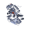
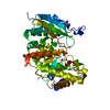
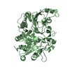
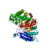
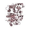
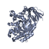
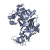
Entry Database : PDB / ID : 4d25Title Crystal structure of the Bombyx mori Vasa helicase (E339Q) in complex with RNA and AMPPNP 5'-R(*UP*GP*AP*CP*AP*UP)-3'BMVLG PROTEIN Keywords / / / Function / homology Function Domain/homology Component
/ / / / / / / / / / / / / / / / / / / / / / / / / / / / / / / Biological species BOMBYX MORI (domestic silkworm)Method / / / Resolution : 1.9 Å Authors Spinelli, P. / Pillai, R.S. / Kadlec, J. / Cusack, S. Journal : Cell(Cambridge,Mass.) / Year : 2014Title : RNA Clamping by Vasa Assembles a Pirna Amplifier Complex on Transposon Transcripts.Authors : Xiol, J. / Spinelli, P. / Laussmann, M.A. / Homolka, D. / Yang, Z. / Cora, E. / Coute, Y. / Conn, S. / Kadlec, J. / Sachidanandam, R. / Kaksonen, M. / Cusack, S. / Ephrussi, A. / Pillai, R.S. History Deposition May 7, 2014 Deposition site / Processing site Revision 1.0 Jun 18, 2014 Provider / Type Revision 1.1 Jul 2, 2014 Group Revision 1.2 Dec 20, 2023 Group Data collection / Database references ... Data collection / Database references / Derived calculations / Other / Refinement description Category chem_comp_atom / chem_comp_bond ... chem_comp_atom / chem_comp_bond / database_2 / pdbx_database_status / pdbx_initial_refinement_model / pdbx_struct_conn_angle / struct_conn / struct_site Item _database_2.pdbx_DOI / _database_2.pdbx_database_accession ... _database_2.pdbx_DOI / _database_2.pdbx_database_accession / _pdbx_database_status.status_code_sf / _pdbx_struct_conn_angle.ptnr1_auth_seq_id / _pdbx_struct_conn_angle.ptnr3_auth_seq_id / _pdbx_struct_conn_angle.value / _struct_conn.pdbx_dist_value / _struct_conn.ptnr2_auth_seq_id / _struct_site.pdbx_auth_asym_id / _struct_site.pdbx_auth_comp_id / _struct_site.pdbx_auth_seq_id
Show all Show less
 Movie
Movie Controller

 Yorodumi
 Open data
 Basic information
 Components
 Keywords
 Function and homology information
 X-RAY DIFFRACTION /
  Authors
 Citation
 Structure visualization
 Downloads & links
 Other downloads
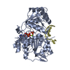


 PDBj
PDBj


 Assembly
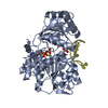


 Mass: 506.196 Da / Num. of mol.: 1 / Source method: obtained synthetically / Formula: C10H17N6O12P3 / Comment: AMP-PNP, energy-carrying molecule analogue*YM
 Mass: 24.305 Da / Num. of mol.: 1 / Source method: obtained synthetically / Formula: Mg
 Mass: 92.094 Da / Num. of mol.: 1 / Source method: obtained synthetically / Formula: C3H8O3
 Sample preparation
 / Beamline: ID14-4 / Wavelength: 0.939
  Processing