regulation of sarcomere organization / MAP kinase scaffold activity / regulation of Rho protein signal transduction / cardiac muscle cell differentiation / regulation of small GTPase mediated signal transduction / RHOB GTPase cycle / NRAGE signals death through JNK / adrenergic receptor signaling pathway / RHOC GTPase cycle / positive regulation of Rho protein signal transduction ...regulation of sarcomere organization / MAP kinase scaffold activity / regulation of Rho protein signal transduction / cardiac muscle cell differentiation / regulation of small GTPase mediated signal transduction / RHOB GTPase cycle / NRAGE signals death through JNK / adrenergic receptor signaling pathway / RHOC GTPase cycle / positive regulation of Rho protein signal transduction / protein kinase A binding / RHOA GTPase cycle / guanyl-nucleotide exchange factor activity / bone development / small GTPase binding / G alpha (12/13) signalling events / heart development / cell cortex / molecular adaptor activity / positive regulation of canonical NF-kappaB signal transduction / G protein-coupled receptor signaling pathway / zinc ion binding / membrane / nucleus / cytosol Similarity search - Function
Protocol: SINGLE WAVELENGTH / Monochromatic (M) / Laue (L): M / Scattering type: x-ray
Radiation wavelength
Wavelength: 0.9763 Å / Relative weight: 1
Reflection
Resolution: 2.75→42.13 Å / Num. obs: 14660 / % possible obs: 100 % / Observed criterion σ(I): 0 / Redundancy: 6.5 % / Rmerge(I) obs: 0.21 / Net I/σ(I): 7.2
Reflection shell
Resolution: 2.75→2.9 Å / Redundancy: 6.5 % / Rmerge(I) obs: 1.18 / Mean I/σ(I) obs: 2.1 / % possible all: 100
-
Processing
Software
Name
Version
Classification
REFMAC
5.8.0069
refinement
MOSFLM
datareduction
Aimless
datascaling
PHASER
phasing
Refinement
Method to determine structure: MOLECULAR REPLACEMENT / Resolution: 2.75→71.55 Å / Cor.coef. Fo:Fc: 0.918 / Cor.coef. Fo:Fc free: 0.84 / SU B: 41.167 / SU ML: 0.361 / Cross valid method: THROUGHOUT / ESU R Free: 0.445 / Stereochemistry target values: MAXIMUM LIKELIHOOD / Details: HYDROGENS HAVE BEEN ADDED IN THE RIDING POSITIONS.
Rfactor
Num. reflection
% reflection
Selection details
Rfree
0.30849
711
4.9 %
RANDOM
Rwork
0.23641
-
-
-
obs
0.23969
13897
99.87 %
-
Solvent computation
Ion probe radii: 0.8 Å / Shrinkage radii: 0.8 Å / VDW probe radii: 1.2 Å / Solvent model: MASK
 Movie
Movie Controller


 Open data
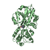
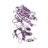
 Basic information
 Components
 Keywords
 Function and homology information
 HOMO SAPIENS (human)
 X-RAY DIFFRACTION /
  Authors
 Citation
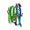
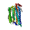
 Structure visualization
 Downloads & links
 Other downloads
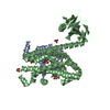
 PDBj
PDBj


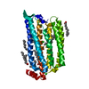
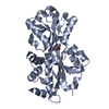
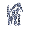
 Assembly


 Sample preparation
 / Beamline: I03 / Wavelength: 0.9763
  Processing