 Movie
Movie Controller

[English] 日本語
 Yorodumi
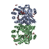
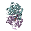
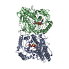
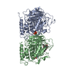
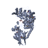
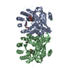
- PDB-4czb: Structure of the sodium proton antiporter MjNhaP1 from Methanocal... -
+ Open data

- Basic information
| Entry | Database: PDB / ID: 4czb | ||||||
|---|---|---|---|---|---|---|---|
| Title | Structure of the sodium proton antiporter MjNhaP1 from Methanocaldococcus jannaschii at pH 8. | ||||||
 Components Components | (NA(+)/H(+) ANTIPORTER 1) x 2 | ||||||
 Keywords Keywords | MEMBRANE PROTEIN / ANTIPORTER / TRANSPORTER / EXCHANGER / CPA | ||||||
| Function / homology |  Function and homology information Function and homology informationpotassium:proton antiporter activity / sodium ion transport / intracellular potassium ion homeostasis / identical protein binding / plasma membrane Similarity search - Function | ||||||
| Biological species |   METHANOCALDOCOCCUS JANNASCHII (archaea) METHANOCALDOCOCCUS JANNASCHII (archaea) | ||||||
| Method |  X-RAY DIFFRACTION / SYNCHROTRON / MOLECULAR REPLACEMENT / Resolution: 3.5 Å X-RAY DIFFRACTION / SYNCHROTRON / MOLECULAR REPLACEMENT / Resolution: 3.5 Å | ||||||
 Authors Authors | Woehlert, D. / Paulino, C. / Kapotova, E. / Kuhlbrandt, W. / Yildiz, O. | ||||||
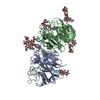
 Citation Citation | Journal: Elife / Year: 2014 Title: Structure and transport mechanism of the sodium/proton antiporter MjNhaP1. Authors: Cristina Paulino / David Wöhlert / Ekaterina Kapotova / Özkan Yildiz / Werner Kühlbrandt /  Abstract: Sodium/proton antiporters are essential for sodium and pH homeostasis and play a major role in human health and disease. We determined the structures of the archaeal sodium/proton antiporter MjNhaP1 ...Sodium/proton antiporters are essential for sodium and pH homeostasis and play a major role in human health and disease. We determined the structures of the archaeal sodium/proton antiporter MjNhaP1 in two complementary states. The inward-open state was obtained by x-ray crystallography in the presence of sodium at pH 8, where the transporter is highly active. The outward-open state was obtained by electron crystallography without sodium at pH 4, where MjNhaP1 is inactive. Comparison of both structures reveals a 7° tilt of the 6 helix bundle. (22)Na(+) uptake measurements indicate non-cooperative transport with an activity maximum at pH 7.5. We conclude that binding of a Na(+) ion from the outside induces helix movements that close the extracellular cavity, open the cytoplasmic funnel, and result in a ∼5 Å vertical relocation of the ion binding site to release the substrate ion into the cytoplasm. | ||||||
| History |
|
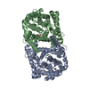
- Structure visualization
| Structure viewer | Molecule: MolmilJmol/JSmol |
|---|
- Downloads & links
-Download
| PDBx/mmCIF format | 4czb.cif.gz | 312.3 KB | Display | PDBx/mmCIF format |
|---|---|---|---|---|
| PDB format | pdb4czb.ent.gz | 256.1 KB | Display | PDB format |
| PDBx/mmJSON format | 4czb.json.gz | Tree view | PDBx/mmJSON format | |
| Others |  Other downloads Other downloads |
-Validation report
| Arichive directory | https://data.pdbj.org/pub/pdb/validation_reports/cz/4czbftp://data.pdbj.org/pub/pdb/validation_reports/cz/4czb | HTTPS FTP |
|---|
-Related structure data
| Related structure data |  2636C  4d0aC 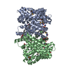 4cz8S S: Starting model for refinement C: citing same article ( |
|---|---|
| Similar structure data |


-Links
 PDBj
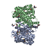
PDBj- Assembly
| Deposited unit | 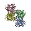
| ||||||||
|---|---|---|---|---|---|---|---|---|---|
| 1 | 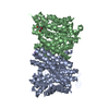
| ||||||||
| 2 | 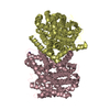
| ||||||||
| Unit cell |
|
-Components
| #1: Protein | Mass: 45998.891 Da / Num. of mol.: 3 / Fragment: TRANSPORTER DOMAIN, RESIDUES 1-426 Source method: isolated from a genetically manipulated source Source: (gene. exp.) METHANOCALDOCOCCUS JANNASCHII (archaea)Production host:  #2: Protein | | Mass: 46012.918 Da / Num. of mol.: 1 / Fragment: TRANSPORTER DOMAIN, RESIDUES 1-426 Source method: isolated from a genetically manipulated source Source: (gene. exp.) METHANOCALDOCOCCUS JANNASCHII (archaea)Production host: #3: Chemical |   Mass: 39.098 Da / Num. of mol.: 2 / Source method: obtained synthetically / Formula: K Mass: 39.098 Da / Num. of mol.: 2 / Source method: obtained synthetically / Formula: K#4: Chemical | ChemComp-TAM / | 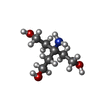  Mass: 163.215 Da / Num. of mol.: 1 / Source method: obtained synthetically / Formula: C7H17NO3 / Comment: pH buffer*YM Mass: 163.215 Da / Num. of mol.: 1 / Source method: obtained synthetically / Formula: C7H17NO3 / Comment: pH buffer*YM |
|---|
-Experimental details
-Experiment
| Experiment | Method: X-RAY DIFFRACTION / Number of used crystals: 1 |
|---|
- Sample preparation
| Crystal | Density Matthews: 3.49 Å3/Da / Density % sol: 61 % / Description: NONE |
|---|---|
| Crystal grow | pH: 8.2 / Details: pH 8.2 |
-Data collection
| Diffraction | Mean temperature: 100 K |
|---|---|
| Diffraction source | Source: SYNCHROTRON / Site: ESRF  / Beamline: ID23-1 / Wavelength: 0.9763 / Beamline: ID23-1 / Wavelength: 0.9763 |
| Detector | Date: Jul 13, 2012 |
| Radiation | Protocol: SINGLE WAVELENGTH / Monochromatic (M) / Laue (L): M / Scattering type: x-ray |
| Radiation wavelength | Wavelength: 0.9763 Å / Relative weight: 1 |
| Reflection | Resolution: 3.5→32 Å / Num. obs: 58249 / % possible obs: 99.3 % / Observed criterion σ(I): 1.7 / Redundancy: 7 % / Rmerge(I) obs: 0.09 / Net I/σ(I): 6 |
| Reflection shell | Resolution: 3.5→3.72 Å / Redundancy: 7.2 % / Rmerge(I) obs: 0.57 / Mean I/σ(I) obs: 1.7 / % possible all: 99.2 |
- Processing
| Software |
| ||||||||||||||||
|---|---|---|---|---|---|---|---|---|---|---|---|---|---|---|---|---|---|
| Refinement | Method to determine structure: MOLECULAR REPLACEMENT Starting model: PDB ENTRY 4CZ8 Resolution: 3.5→32 Å / σ(F): 6
| ||||||||||||||||
| Solvent computation | Shrinkage radii: 0.9 Å / VDW probe radii: 1.11 Å / Solvent model: FLAT BULK SOLVENT MODEL | ||||||||||||||||
| Refinement step | Cycle: LAST / Resolution: 3.5→32 Å
|
