 Movie
Movie Controller

[English] 日本語
 Yorodumi
- EMDB-2636: 3D EM map of the sodium proton antiporter MjNhaP1 from Methanocal... -
+ Open data

- Basic information
| Entry | Database: EMDB / ID: EMD-2636 | |||||||||
|---|---|---|---|---|---|---|---|---|---|---|
| Title | 3D EM map of the sodium proton antiporter MjNhaP1 from Methanocaldococcus jannaschii | |||||||||
 Map data Map data | A B-factor of -200 was applied. | |||||||||
 Sample Sample |
| |||||||||
 Keywords Keywords | membrane protein / antiporter / transporter / exchanger / CPA | |||||||||
| Function / homology |  Function and homology information Function and homology information: / transport / potassium:proton antiporter activity / antiporter activity / sodium ion transport / intracellular potassium ion homeostasis / membrane => GO:0016020 / monoatomic cation transport / monoatomic ion transport / proton transmembrane transport ...: / transport / potassium:proton antiporter activity / antiporter activity / sodium ion transport / intracellular potassium ion homeostasis / membrane => GO:0016020 / monoatomic cation transport / monoatomic ion transport / proton transmembrane transport / transmembrane transport / membrane / identical protein binding / plasma membrane Similarity search - Function | |||||||||
| Biological species |   Methanocaldococcus jannaschii (archaea) Methanocaldococcus jannaschii (archaea) | |||||||||
| Method | electron crystallography / cryo EM / negative staining / Resolution: 6.0 Å | |||||||||
 Authors Authors | Paulino C / Woehlert D / Yildiz O / Kuhlbrandt W | |||||||||
 Citation Citation | Journal: Elife / Year: 2014 Title: Structure and transport mechanism of the sodium/proton antiporter MjNhaP1. Authors: Cristina Paulino / David Wöhlert / Ekaterina Kapotova / Özkan Yildiz / Werner Kühlbrandt /  Abstract: Sodium/proton antiporters are essential for sodium and pH homeostasis and play a major role in human health and disease. We determined the structures of the archaeal sodium/proton antiporter MjNhaP1 ...Sodium/proton antiporters are essential for sodium and pH homeostasis and play a major role in human health and disease. We determined the structures of the archaeal sodium/proton antiporter MjNhaP1 in two complementary states. The inward-open state was obtained by x-ray crystallography in the presence of sodium at pH 8, where the transporter is highly active. The outward-open state was obtained by electron crystallography without sodium at pH 4, where MjNhaP1 is inactive. Comparison of both structures reveals a 7° tilt of the 6 helix bundle. (22)Na(+) uptake measurements indicate non-cooperative transport with an activity maximum at pH 7.5. We conclude that binding of a Na(+) ion from the outside induces helix movements that close the extracellular cavity, open the cytoplasmic funnel, and result in a ∼5 Å vertical relocation of the ion binding site to release the substrate ion into the cytoplasm. | |||||||||
| History |
|
- Structure visualization
| Movie |
Movie viewer |
|---|---|
| Structure viewer | EM map: SurfViewMolmilJmol/JSmol |
| Supplemental images |

- Downloads & links
-EMDB archive
| Map data | emd_2636.map.gz | 6 MB | EMDB map data format | |
|---|---|---|---|---|
| Header (meta data) | emd-2636-v30.xmlemd-2636.xml | 14.6 KB 14.6 KB | Display Display | EMDB header |
| Images | 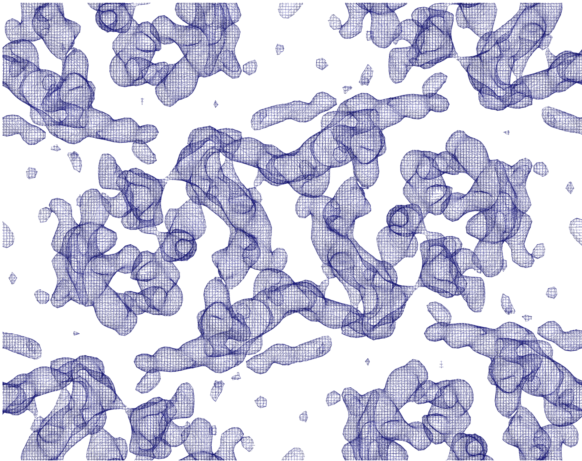 emd_2636.png emd_2636.png | 4 MB | ||
| Archive directory |  http://ftp.pdbj.org/pub/emdb/structures/EMD-2636ftp://ftp.pdbj.org/pub/emdb/structures/EMD-2636 http://ftp.pdbj.org/pub/emdb/structures/EMD-2636ftp://ftp.pdbj.org/pub/emdb/structures/EMD-2636 | HTTPS FTP |
-Related structure data
| Related structure data | 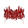 4d0aMC 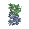 4czbC M: atomic model generated by this map C: citing same article ( |
|---|---|
| Similar structure data |
-Links
| EMDB pages | EMDB (EBI/PDBe) / EMDataResource |
|---|
-Map
| File | Download / File: emd_2636.map.gz / Format: CCP4 / Size: 6.3 MB / Type: IMAGE STORED AS FLOATING POINT NUMBER (4 BYTES) | ||||||||||||||||||||||||||||||||||||||||||||||||||||||||||||||||||||
|---|---|---|---|---|---|---|---|---|---|---|---|---|---|---|---|---|---|---|---|---|---|---|---|---|---|---|---|---|---|---|---|---|---|---|---|---|---|---|---|---|---|---|---|---|---|---|---|---|---|---|---|---|---|---|---|---|---|---|---|---|---|---|---|---|---|---|---|---|---|
| Annotation | A B-factor of -200 was applied. | ||||||||||||||||||||||||||||||||||||||||||||||||||||||||||||||||||||
| Projections & slices | Image control
Images are generated by Spider. generated in cubic-lattice coordinate | ||||||||||||||||||||||||||||||||||||||||||||||||||||||||||||||||||||
| Voxel size | X: 1.01875 Å / Y: 0.99327 Å / Z: 1 Å | ||||||||||||||||||||||||||||||||||||||||||||||||||||||||||||||||||||
| Density |
| ||||||||||||||||||||||||||||||||||||||||||||||||||||||||||||||||||||
| Symmetry | Space group: 18 | ||||||||||||||||||||||||||||||||||||||||||||||||||||||||||||||||||||
| Details | EMDB XML:
CCP4 map header:
| ||||||||||||||||||||||||||||||||||||||||||||||||||||||||||||||||||||
 Y (Sec.)
 X (Row.)
 Z (Col.)


-Supplemental data
- Sample components
-Entire : 3D EM map of the sodium/proton antiporter MjNhaP1
| Entire | Name: 3D EM map of the sodium/proton antiporter MjNhaP1 |
|---|---|
| Components |
|
-Supramolecule #1000: 3D EM map of the sodium/proton antiporter MjNhaP1
| Supramolecule | Name: 3D EM map of the sodium/proton antiporter MjNhaP1 / type: sample / ID: 1000 / Details: protein was purified in absence of sodium. / Oligomeric state: Dimer / Number unique components: 1 |
|---|---|
| Molecular weight | Experimental: 46 KDa / Theoretical: 46 KDa / Method: SDS-PAGE, MS |
-Macromolecule #1: MjNhaP1
| Macromolecule | Name: MjNhaP1 / type: protein_or_peptide / ID: 1 / Name.synonym: NhaP1 / Number of copies: 2 / Oligomeric state: Dimer / Recombinant expression: Yes |
|---|---|
| Source (natural) | Organism: Methanocaldococcus jannaschii (archaea) / synonym: Methanocaldococcus jannaschii / Location in cell: Plasma membrane |
| Molecular weight | Experimental: 46 KDa / Theoretical: 46 KDa |
| Recombinant expression | Organism:  |
| Sequence | UniProtKB: Na(+)/H(+) antiporter 1 GO: transport, monoatomic ion transport, monoatomic cation transport, sodium ion transport, transmembrane transport, proton transmembrane transport, antiporter activity, GO: 0015299, identical ...GO: transport, monoatomic ion transport, monoatomic cation transport, sodium ion transport, transmembrane transport, proton transmembrane transport, antiporter activity, GO: 0015299, identical protein binding, plasma membrane, membrane, membrane => GO:0016020 InterPro: Cation/H+ exchanger |
-Experimental details
-Structure determination
| Method | negative staining, cryo EM |
|---|---|
 Processing Processing | electron crystallography |
| Aggregation state | 2D array |
-Sample preparation
| Concentration | 1 mg/mL |
|---|---|
| Buffer | pH: 4 / Details: 25mM KAc pH4, 200mM KCl, 5mM glycerol, 5mM MPD |
| Staining | Type: NEGATIVE / Details: back injection method with 4% trehalose |
| Grid | Details: 400 mesh copper grid |
| Vitrification | Cryogen name: NITROGEN / Chamber humidity: 20 % / Chamber temperature: 77 K / Instrument: OTHER / Details: all buffers used were sodium-free Timed resolved state: sample was plunge-frozen in liquid nitrogen Method: back injection method (Wang & Kuhlbrandt, 1991) with 4% trehalose |
| Details | E.coli polar lipids with a lipid-to-protein ration (LPR) of 0.4-0.5 were used. 2D crystals were grown by slow removal of detergent (0.15% DM) by dialysis. |
| Crystal formation | Details: E.coli polar lipids with a lipid-to-protein ration (LPR) of 0.4-0.5 were used. 2D crystals were grown by slow removal of detergent (0.15% DM) by dialysis. |
- Electron microscopy
| Microscope | JEOL 3000SFF |
|---|---|
| Temperature | Min: 4 K / Max: 10 K / Average: 4 K |
| Alignment procedure | Legacy - Astigmatism: objective lens was corrected at 60kx and/or 300kx magnification Legacy - Electron beam tilt params: - |
| Specialist optics | Energy filter - Name: - |
| Date | Dec 1, 2012 |
| Image recording | Category: FILM / Film or detector model: KODAK SO-163 FILM / Digitization - Scanner: ZEISS SCAI / Digitization - Sampling interval: 7 µm / Number real images: 128 / Average electron dose: 25 e/Å2 |
| Tilt angle min | 0 |
| Electron beam | Acceleration voltage: 300 kV / Electron source:  FIELD EMISSION GUN FIELD EMISSION GUN |
| Electron optics | Calibrated magnification: 53000 / Illumination mode: SPOT SCAN / Imaging mode: OTHER / Cs: 1.6 mm / Nominal defocus max: 1.8 µm / Nominal defocus min: 0.12 µm / Nominal magnification: 60000 |
| Sample stage | Specimen holder: helium-cooled top entry stage with fixed specimen holder. Specimen holder model: JEOL / Tilt angle max: 45 / Tilt series - Axis1 - Min angle: 0 ° / Tilt series - Axis1 - Max angle: 45 ° |
-Image processing
| Details | Images were processed with the 2dx software. |
|---|---|
| Final reconstruction | Resolution.type: BY AUTHOR / Resolution: 6.0 Å / Resolution method: OTHER / Software - Name: 2dx Details: 6A in plane resolution and 14A resolution in the z direction. |
| Crystal parameters | Unit cell - A: 81.5 Å / Unit cell - B: 103.3 Å / Unit cell - C: 200 Å / Unit cell - γ: 90.0 ° / Unit cell - α: 90.0 ° / Unit cell - β: 90.0 ° / Plane group: P 2 21 21 |
| CTF correction | Details: 2dx |
-Atomic model buiding 1
| Initial model | PDB ID: Chain - Chain ID: B |
|---|---|
| Software | Name: Coot |
| Details | The X-ray structure of the same protein (4czb) obtained at different conditions was manually fitted to the 3D EM density map. |
| Refinement | Space: REAL / Protocol: RIGID BODY FIT |
| Output model | PDB-4d0a: |
