 Movie
Movie Controller


+ Open data

- Basic information
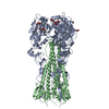
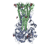
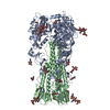
| Entry | Database: PDB / ID: 4cqz | |||||||||
|---|---|---|---|---|---|---|---|---|---|---|
| Title | Crystal Structure of H5 (VN1194) Gln196Arg Mutant Haemagglutinin | |||||||||
 Components Components | (Hemagglutinin ...) x 2 | |||||||||
 Keywords Keywords | VIRAL PROTEIN / HAEMAGGLUTININ MUTANT / SIALIC ACID / GLYCOPROTEIN / VIRUS RECEPTOR / AVIAN FLU / SIALYLLACTOSAMINE / 3SLN / 3'SLN / 6SLN / 6'SLN / LSTA | |||||||||
| Function / homology |  Function and homology information Function and homology informationclathrin-dependent endocytosis of virus by host cell / host cell surface receptor binding / fusion of virus membrane with host plasma membrane / fusion of virus membrane with host endosome membrane / viral envelope / virion attachment to host cell / host cell plasma membrane / virion membrane Similarity search - Function | |||||||||
| Biological species |   Influenza A virus Influenza A virus | |||||||||
| Method |  X-RAY DIFFRACTION / MOLECULAR REPLACEMENT / Resolution: 2.7 Å X-RAY DIFFRACTION / MOLECULAR REPLACEMENT / Resolution: 2.7 Å | |||||||||
 Authors Authors | Liu, J. / Xiong, X. / Xiao, H. / Martin, S.R. / Coombs, P.J. / Collins, P.J. / Vachieri, S.G. / Walker, P.A. / Lin, Y.P. / McCauley, J.W. ...Liu, J. / Xiong, X. / Xiao, H. / Martin, S.R. / Coombs, P.J. / Collins, P.J. / Vachieri, S.G. / Walker, P.A. / Lin, Y.P. / McCauley, J.W. / Gamblin, S.J. / Skehel, J.J. | |||||||||
 Citation Citation | Journal: Virology / Year: 2014 Title: Enhanced Human Receptor Binding by H5 Haemagglutinins. Authors: Xiong, X. / Xiao, H. / Martin, S.R. / Coombs, P.J. / Liu, J. / Collins, P.J. / Vachieri, S.G. / Walker, P.A. / Lin, Y.P. / Mccauley, J.W. / Gamblin, S.J. / Skehel, J.J. | |||||||||
| History |
|
- Structure visualization
| Structure viewer | Molecule: MolmilJmol/JSmol |
|---|
- Downloads & links
-Download
| PDBx/mmCIF format | 4cqz.cif.gz | 213.3 KB | Display | PDBx/mmCIF format |
|---|---|---|---|---|
| PDB format | pdb4cqz.ent.gz | 173.5 KB | Display | PDB format |
| PDBx/mmJSON format | 4cqz.json.gz | Tree view | PDBx/mmJSON format | |
| Others |  Other downloads Other downloads |
-Validation report
| Arichive directory | https://data.pdbj.org/pub/pdb/validation_reports/cq/4cqzftp://data.pdbj.org/pub/pdb/validation_reports/cq/4cqz | HTTPS FTP |
|---|
-Related structure data
| Related structure data | 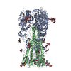 4cqpC 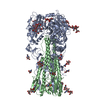 4cqqC 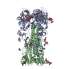 4cqrC  4cqsC  4cquC  4cqvC  4cqwC  4cqxC  4cqyC  4cr0C  5ajmC  4bgwS C: citing same article ( S: Starting model for refinement |
|---|---|
| Similar structure data |

-Links
 PDBj
PDBj


- Assembly
| Deposited unit | 
| ||||||||
|---|---|---|---|---|---|---|---|---|---|
| 1 | 
| ||||||||
| Unit cell |
|
-Components
-Hemagglutinin ... , 2 types, 2 molecules AB
| #1: Protein | Mass: 36979.828 Da / Num. of mol.: 1 Fragment: HA1 OF TRYPSIN RELEASED ECTODOMAIN, RESIDUES 17-340 Mutation: Q196R Source method: isolated from a genetically manipulated source Source: (gene. exp.) Influenza A virus / Gene: HA / Variant: GLN196ARG MUTANT / Production host:  |
|---|---|
| #2: Protein | Mass: 19097.990 Da / Num. of mol.: 1 Fragment: HA2 OF TRYPSIN RELEASED ECTODOMAIN, RESIDUES 347-512 Source method: isolated from a genetically manipulated source Source: (gene. exp.) Influenza A virus / Gene: HA / Production host: |
-Sugars , 4 types, 4 molecules 
| #3: Polysaccharide | N-acetyl-alpha-neuraminic acid-(2-3)-beta-D-galactopyranose-(1-3)-2-acetamido-2-deoxy-beta-D-glucopyranose Source method: isolated from a genetically manipulated source |
|---|---|
| #4: Polysaccharide | 2-acetamido-2-deoxy-beta-D-glucopyranose-(1-3)-2-acetamido-2-deoxy-beta-D-glucopyranose Source method: isolated from a genetically manipulated source |
| #5: Polysaccharide | 2-acetamido-2-deoxy-beta-D-glucopyranose-(1-4)-2-acetamido-2-deoxy-beta-D-glucopyranose Source method: isolated from a genetically manipulated source |
| #6: Sugar | ChemComp-NAG /  Type: D-saccharide, beta linking / Mass: 221.208 Da / Num. of mol.: 1 Type: D-saccharide, beta linking / Mass: 221.208 Da / Num. of mol.: 1Source method: isolated from a genetically manipulated source Formula: C8H15NO6 |
-Non-polymers , 2 types, 88 molecules 

| #7: Chemical | ChemComp-MPO /  Mass: 209.263 Da / Num. of mol.: 1 / Source method: obtained synthetically / Formula: C7H15NO4S / Comment: pH buffer*YM Mass: 209.263 Da / Num. of mol.: 1 / Source method: obtained synthetically / Formula: C7H15NO4S / Comment: pH buffer*YM |
|---|---|
| #8: Water | ChemComp-HOH / Mass: 18.015 Da / Num. of mol.: 87 / Source method: isolated from a natural source / Formula: H2O |
-Details
| Has protein modification | Y |
|---|---|
| Sequence details | MULTIBASIC |
-Experimental details
-Experiment
| Experiment | Method: X-RAY DIFFRACTION / Number of used crystals: 1 |
|---|
- Sample preparation
| Crystal | Density Matthews: 3.74 Å3/Da / Density % sol: 67 % / Description: NONE |
|---|---|
| Crystal grow | Details: 0.1 M HEPES/MOPS PH 7.0, 0.05 M MGCL2, 28-30% PEG 550 MME, SEEDED WITH CRUSHED WILD-TYPE VN1194 HA CRYSTALS. |
-Data collection
| Diffraction | Mean temperature: 100 K |
|---|---|
| Diffraction source | Source: ROTATING ANODE / Wavelength: 1.54 |
| Radiation | Protocol: SINGLE WAVELENGTH / Monochromatic (M) / Laue (L): M / Scattering type: x-ray |
| Radiation wavelength | Wavelength: 1.54 Å / Relative weight: 1 |
| Reflection | Resolution: 2.7→74.65 Å / Num. obs: 24883 / % possible obs: 99.9 % / Observed criterion σ(I): 1.9 / Redundancy: 5.2 % / Rmerge(I) obs: 0.08 / Net I/σ(I): 11.7 |
| Reflection shell | Resolution: 2.7→2.85 Å / Redundancy: 3.3 % / Rmerge(I) obs: 0.43 / Mean I/σ(I) obs: 1.9 / % possible all: 99.4 |
- Processing
| Software |
| ||||||||||||||||||||||||||||||||||||||||||||||||||||||||||||||||||||||||||||||||||||||||||||||||||||||||||||||||||||||||||||||||||||||||||||||||||||||||||||||||||||||||||||||||||||||
|---|---|---|---|---|---|---|---|---|---|---|---|---|---|---|---|---|---|---|---|---|---|---|---|---|---|---|---|---|---|---|---|---|---|---|---|---|---|---|---|---|---|---|---|---|---|---|---|---|---|---|---|---|---|---|---|---|---|---|---|---|---|---|---|---|---|---|---|---|---|---|---|---|---|---|---|---|---|---|---|---|---|---|---|---|---|---|---|---|---|---|---|---|---|---|---|---|---|---|---|---|---|---|---|---|---|---|---|---|---|---|---|---|---|---|---|---|---|---|---|---|---|---|---|---|---|---|---|---|---|---|---|---|---|---|---|---|---|---|---|---|---|---|---|---|---|---|---|---|---|---|---|---|---|---|---|---|---|---|---|---|---|---|---|---|---|---|---|---|---|---|---|---|---|---|---|---|---|---|---|---|---|---|---|
| Refinement | Method to determine structure: MOLECULAR REPLACEMENT Starting model: PDB ENTRY 4BGW Resolution: 2.7→149.3 Å / Cor.coef. Fo:Fc: 0.94 / Cor.coef. Fo:Fc free: 0.926 / SU B: 26.609 / SU ML: 0.255 / Cross valid method: THROUGHOUT / ESU R: 0.45 / ESU R Free: 0.289 / Stereochemistry target values: MAXIMUM LIKELIHOOD / Details: HYDROGENS HAVE BEEN ADDED IN THE RIDING POSITIONS.
| ||||||||||||||||||||||||||||||||||||||||||||||||||||||||||||||||||||||||||||||||||||||||||||||||||||||||||||||||||||||||||||||||||||||||||||||||||||||||||||||||||||||||||||||||||||||
| Solvent computation | Ion probe radii: 0.8 Å / Shrinkage radii: 0.8 Å / VDW probe radii: 1.2 Å / Solvent model: MASK | ||||||||||||||||||||||||||||||||||||||||||||||||||||||||||||||||||||||||||||||||||||||||||||||||||||||||||||||||||||||||||||||||||||||||||||||||||||||||||||||||||||||||||||||||||||||
| Displacement parameters | Biso mean: 74.888 Å2
| ||||||||||||||||||||||||||||||||||||||||||||||||||||||||||||||||||||||||||||||||||||||||||||||||||||||||||||||||||||||||||||||||||||||||||||||||||||||||||||||||||||||||||||||||||||||
| Refinement step | Cycle: LAST / Resolution: 2.7→149.3 Å
| ||||||||||||||||||||||||||||||||||||||||||||||||||||||||||||||||||||||||||||||||||||||||||||||||||||||||||||||||||||||||||||||||||||||||||||||||||||||||||||||||||||||||||||||||||||||
| Refine LS restraints |
|