proton-transporting two-sector ATPase complex, proton-transporting domain / proton-transporting ATP synthase complex / proton-transporting ATP synthase activity, rotational mechanism / lipid binding / plasma membrane Similarity search - Function
F1F0 ATP synthase subunit C / F1FO ATP Synthase / ATP synthase, F0 complex, subunit C, bacterial/chloroplast / ATP synthase, F0 complex, subunit C / F1F0 ATP synthase subunit C superfamily / ATP synthase, F0 complex, subunit C, DCCD-binding site / ATP synthase c subunit signature. / V-ATPase proteolipid subunit C-like domain / F/V-ATP synthase subunit C superfamily / ATP synthase subunit C ...F1F0 ATP synthase subunit C / F1FO ATP Synthase / ATP synthase, F0 complex, subunit C, bacterial/chloroplast / ATP synthase, F0 complex, subunit C / F1F0 ATP synthase subunit C superfamily / ATP synthase, F0 complex, subunit C, DCCD-binding site / ATP synthase c subunit signature. / V-ATPase proteolipid subunit C-like domain / F/V-ATP synthase subunit C superfamily / ATP synthase subunit C / Up-down Bundle / Mainly Alpha Similarity search - Domain/homology
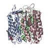
A: ATP SYNTHASE SUBUNIT C B: ATP SYNTHASE SUBUNIT C C: ATP SYNTHASE SUBUNIT C D: ATP SYNTHASE SUBUNIT C E: ATP SYNTHASE SUBUNIT C F: ATP SYNTHASE SUBUNIT C G: ATP SYNTHASE SUBUNIT C H: ATP SYNTHASE SUBUNIT C I: ATP SYNTHASE SUBUNIT C J: ATP SYNTHASE SUBUNIT C K: ATP SYNTHASE SUBUNIT C L: ATP SYNTHASE SUBUNIT C M: ATP SYNTHASE SUBUNIT C hetero molecules
ATPSYNTHASESUBUNITC / ATP SYNTHASE F(0) SECTOR SUBUNIT C / F-TYPE ATPASE SUBUNIT C / F-ATPASE SUBUNIT C / LIPID-BINDING ...ATP SYNTHASE F(0) SECTOR SUBUNIT C / F-TYPE ATPASE SUBUNIT C / F-ATPASE SUBUNIT C / LIPID-BINDING PROTEIN / OF4 C RING
Mass: 6987.461 Da / Num. of mol.: 13 / Source method: isolated from a natural source / Source: (natural) BACILLUS PSEUDOFIRMUS OF4 (bacteria) / References: UniProt: P22483
In the structure databanks used in Yorodumi, some data are registered as the other names, "COVID-19 virus" and "2019-nCoV". Here are the details of the virus and the list of structure data.
Jan 31, 2019. EMDB accession codes are about to change! (news from PDBe EMDB page)
EMDB accession codes are about to change! (news from PDBe EMDB page)
The allocation of 4 digits for EMDB accession codes will soon come to an end. Whilst these codes will remain in use, new EMDB accession codes will include an additional digit and will expand incrementally as the available range of codes is exhausted. The current 4-digit format prefixed with “EMD-” (i.e. EMD-XXXX) will advance to a 5-digit format (i.e. EMD-XXXXX), and so on. It is currently estimated that the 4-digit codes will be depleted around Spring 2019, at which point the 5-digit format will come into force.
The EM Navigator/Yorodumi systems omit the EMD- prefix.
Related info.:Q: What is EMD? / ID/Accession-code notation in Yorodumi/EM Navigator
Yorodumi is a browser for structure data from EMDB, PDB, SASBDB, etc.
This page is also the successor to EM Navigator detail page, and also detail information page/front-end page for Omokage search.
The word "yorodu" (or yorozu) is an old Japanese word meaning "ten thousand". "mi" (miru) is to see.
Related info.:EMDB / PDB / SASBDB / Comparison of 3 databanks / Yorodumi Search / Aug 31, 2016. New EM Navigator & Yorodumi / Yorodumi Papers / Jmol/JSmol / Function and homology information / Changes in new EM Navigator and Yorodumi
 Movie
Movie Controller

 Yorodumi
 Open data
 Basic information
 Components
 Keywords
 Function and homology information
 BACILLUS PSEUDOFIRMUS OF4 (bacteria)
 X-RAY DIFFRACTION /
  Authors
 Citation
 Structure visualization
 Downloads & links
 Other downloads


 PDBj
PDBj

 Assembly
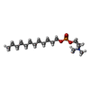
 Mass: 351.462 Da / Num. of mol.: 13 / Source method: obtained synthetically / Formula: C17H38NO4P
 Mass: 22.990 Da / Num. of mol.: 10 / Source method: obtained synthetically / Formula: Na
 Type: D-saccharide / Mass: 510.615 Da / Num. of mol.: 1 / Source method: obtained synthetically / Formula: C24H46O11 / Comment: detergent*YM
 Mass: 18.015 Da / Num. of mol.: 74 / Source method: isolated from a natural source / Formula: H2O
 Sample preparation
 / Beamline: ID23-1 / Wavelength: 0.8551
  Processing