 Movie
Movie Controller

[English] 日本語
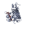
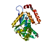
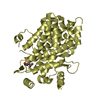
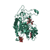
 Yorodumi
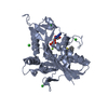
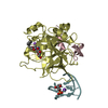
- PDB-4c9q: Structure of yeast mitochondrial ADP/ATP carrier isoform 3 inhibi... -
+ Open data

- Basic information
| Entry | Database: PDB / ID: 4c9q | ||||||
|---|---|---|---|---|---|---|---|
| Title | Structure of yeast mitochondrial ADP/ATP carrier isoform 3 inhibited by carboxyatractyloside (P21 crystal form) | ||||||
 Components Components | ADP, ATP CARRIER PROTEIN 3 | ||||||
 Keywords Keywords | TRANSPORT PROTEIN / MITOCHONDRIAL CARRIER / ADP/ATP CARRIER | ||||||
| Function / homology |  Function and homology information Function and homology informationTransport of nucleosides and free purine and pyrimidine bases across the plasma membrane / mitochondrial ADP transmembrane transport / Mitochondrial protein import / ATP:ADP antiporter activity / mitochondrial ATP transmembrane transport / heme transport / Mitochondrial protein degradation / anaerobic respiration / transmembrane transport / mitochondrial inner membrane ...Transport of nucleosides and free purine and pyrimidine bases across the plasma membrane / mitochondrial ADP transmembrane transport / Mitochondrial protein import / ATP:ADP antiporter activity / mitochondrial ATP transmembrane transport / heme transport / Mitochondrial protein degradation / anaerobic respiration / transmembrane transport / mitochondrial inner membrane / mitochondrion / identical protein binding Similarity search - Function | ||||||
| Biological species |  | ||||||
| Method |  X-RAY DIFFRACTION / SYNCHROTRON / MOLECULAR REPLACEMENT / Resolution: 3.2 Å X-RAY DIFFRACTION / SYNCHROTRON / MOLECULAR REPLACEMENT / Resolution: 3.2 Å | ||||||
 Authors Authors | Ruprecht, J.J. / Hellawell, A.M. / Harding, M. / Crichton, P.G. / McCoy, A.J. / Kunji, E.R.S. | ||||||
 Citation Citation | Journal: Proc.Natl.Acad.Sci.USA / Year: 2014 Title: Structures of Yeast Mitochondrial Adp/ATP Carriers Support a Domain-Based Alternating-Access Transport Mechanism Authors: Ruprecht, J.J. / Hellawell, A.M. / Harding, M. / Crichton, P.G. / Mccoy, A.J. / Kunji, E.R.S. | ||||||
| History |
|
- Structure visualization
| Structure viewer | Molecule: MolmilJmol/JSmol |
|---|
- Downloads & links
-Download
| PDBx/mmCIF format | 4c9q.cif.gz | 133.5 KB | Display | PDBx/mmCIF format |
|---|---|---|---|---|
| PDB format | pdb4c9q.ent.gz | 100.1 KB | Display | PDB format |
| PDBx/mmJSON format | 4c9q.json.gz | Tree view | PDBx/mmJSON format | |
| Others |  Other downloads Other downloads |
-Validation report
| Arichive directory | https://data.pdbj.org/pub/pdb/validation_reports/c9/4c9qftp://data.pdbj.org/pub/pdb/validation_reports/c9/4c9q | HTTPS FTP |
|---|
-Related structure data
| Related structure data |  4c9gC 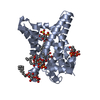 4c9hC 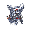 4c9jSC C: citing same article ( S: Starting model for refinement |
|---|---|
| Similar structure data |


-Links
 PDBj
PDBj
- Assembly
| Deposited unit | 
| ||||||||
|---|---|---|---|---|---|---|---|---|---|
| 1 | 
| ||||||||
| 2 | 
| ||||||||
| Unit cell |
| ||||||||
| Noncrystallographic symmetry (NCS) | NCS oper: (Code: given Matrix: (0.99748, -0.06686, -0.02364), Vector: |
-Components
| #1: Protein | Mass: 35255.980 Da / Num. of mol.: 2 / Fragment: RESIDUES 3-307 Source method: isolated from a genetically manipulated source Source: (gene. exp.) Production host: #2: Chemical |   Mass: 770.816 Da / Num. of mol.: 2 / Source method: obtained synthetically / Formula: C31H46O18S2 Mass: 770.816 Da / Num. of mol.: 2 / Source method: obtained synthetically / Formula: C31H46O18S2#3: Chemical | ChemComp-CDL /   Mass: 1464.043 Da / Num. of mol.: 6 / Source method: obtained synthetically / Formula: C81H156O17P2 / Comment: phospholipid*YM Mass: 1464.043 Da / Num. of mol.: 6 / Source method: obtained synthetically / Formula: C81H156O17P2 / Comment: phospholipid*YM |
|---|
-Experimental details
-Experiment
| Experiment | Method: X-RAY DIFFRACTION / Number of used crystals: 1 |
|---|
- Sample preparation
| Crystal | Density Matthews: 2.3 Å3/Da / Density % sol: 46.8 % / Description: NONE |
|---|---|
| Crystal grow | pH: 8.5 Details: 0.1M TRIS PH 8.5, 5MM SODIUM CITRATE, 11% T-BUTANOL, 26% PEG400 |
-Data collection
| Diffraction | Mean temperature: 100 K |
|---|---|
| Diffraction source | Source: SYNCHROTRON / Site: ESRF  / Beamline: ID23-2 / Wavelength: 0.8726 / Beamline: ID23-2 / Wavelength: 0.8726 |
| Detector | Type: MARRESEARCH / Detector: CCD / Date: Mar 12, 2010 |
| Radiation | Protocol: SINGLE WAVELENGTH / Monochromatic (M) / Laue (L): M / Scattering type: x-ray |
| Radiation wavelength | Wavelength: 0.8726 Å / Relative weight: 1 |
| Reflection | Resolution: 3.2→41.45 Å / Num. obs: 11131 / % possible obs: 98 % / Observed criterion σ(I): 6 / Redundancy: 2.1 % / Biso Wilson estimate: 45.02 Å2 / Rmerge(I) obs: 0.13 / Net I/σ(I): 5.7 |
| Reflection shell | Resolution: 3.2→3.42 Å / Redundancy: 2.1 % / Rmerge(I) obs: 0.41 / Mean I/σ(I) obs: 2.5 / % possible all: 98.3 |
- Processing
| Software |
| |||||||||||||||||||||||||||||||||||
|---|---|---|---|---|---|---|---|---|---|---|---|---|---|---|---|---|---|---|---|---|---|---|---|---|---|---|---|---|---|---|---|---|---|---|---|---|
| Refinement | Method to determine structure: MOLECULAR REPLACEMENT Starting model: PDB ENTRY 4C9J Resolution: 3.2→41.449 Å / SU ML: 0.44 / σ(F): 1.38 / Phase error: 34.34 / Stereochemistry target values: ML Details: RESIDUES 151-155 AND 207-215 OF CHAIN A, AND 149-155 AND 208-214 OF CHAIN B, ARE UNMODELLED OWING TO POOR DENSITY
| |||||||||||||||||||||||||||||||||||
| Solvent computation | Shrinkage radii: 0.9 Å / VDW probe radii: 1.11 Å / Solvent model: FLAT BULK SOLVENT MODEL | |||||||||||||||||||||||||||||||||||
| Refinement step | Cycle: LAST / Resolution: 3.2→41.449 Å
| |||||||||||||||||||||||||||||||||||
| Refine LS restraints |
| |||||||||||||||||||||||||||||||||||
| LS refinement shell |
|
